Frontal Eye Fields — Function, Location & Quiz
The frontal eye fields convert a visual or attentional decision into a voluntary saccade. They are not the brainstem machinery that moves the eyes; they help select the target and send the command toward the superior colliculus and gaze generators.
Their neurons also reveal decision formation before movement begins, with activity rising as evidence accumulates. That makes this small frontal region a useful bridge between cognitive choice, spatial attention and a measurable motor act.
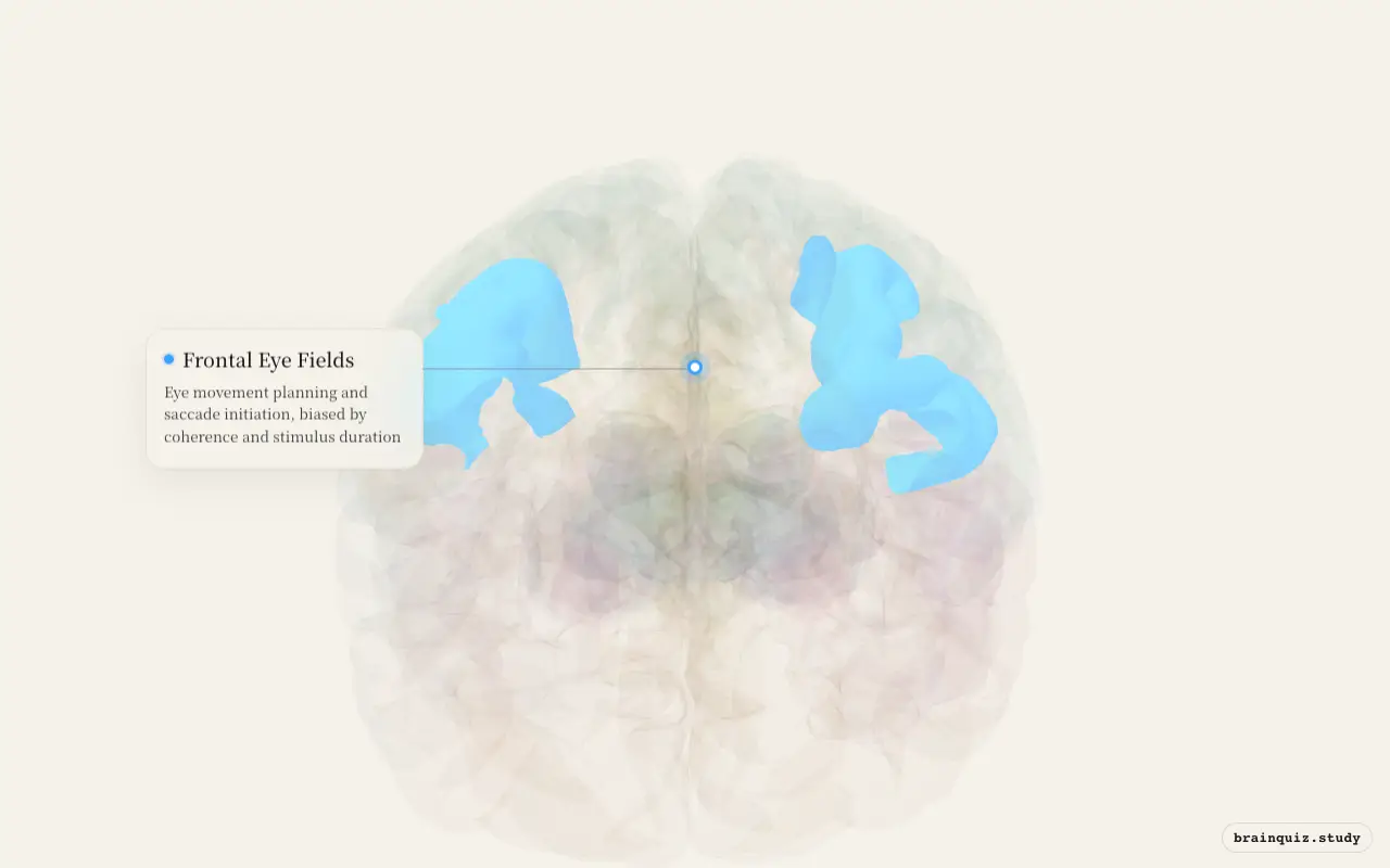
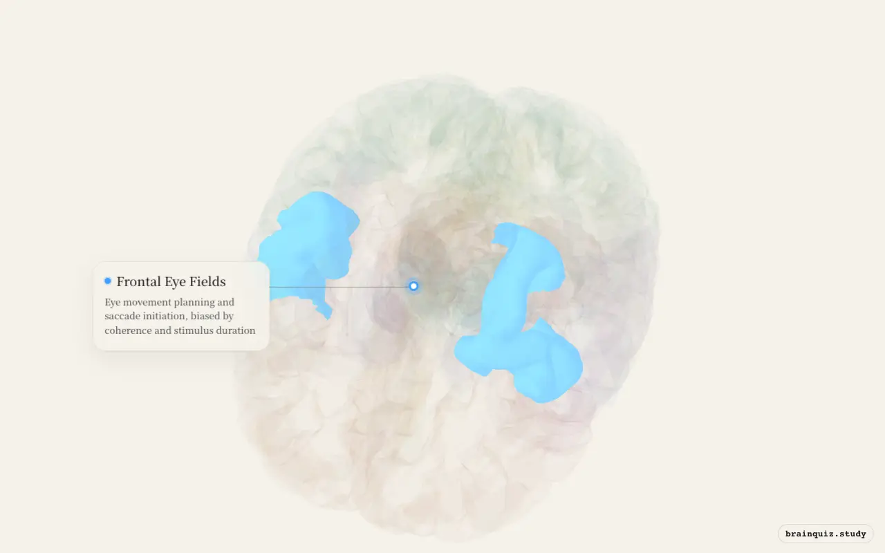
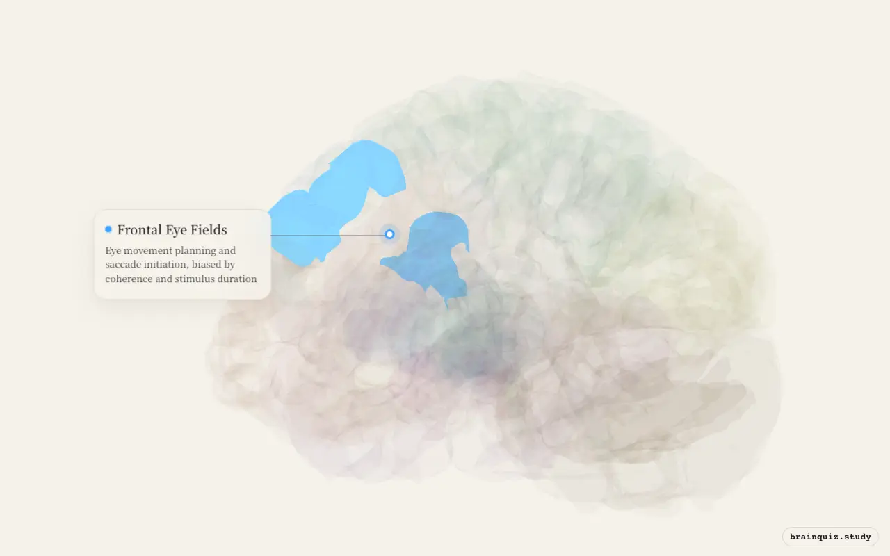
Test this anatomy in the Lesion Localization Quiz: 15 Clinical Questions or explore it on the interactive 3D brain.
Where is the Frontal Eye Fields located?
Eye movement planning and saccade initiation, biased by coherence and stimulus duration.
What does the Frontal Eye Fields do?
- Eye movement planning — voluntary saccades
- Evidence accumulation — graded neural response biased by coherence and duration
- Attention shifts via planned eye movements
- Neurons respond to both movement direction and reward value
Frontal Eye Fields pathways and connections
- FEF → Superior colliculus (saccade execution)
- FEF ← Parietal cortex (spatial attention input)
- FEF ↔ Basal ganglia (gating via SNr — eye movement circuit)
What happens when the Frontal Eye Fields is damaged?
- Lesion impairs voluntary saccades (can't look where you want)
- Stimulation evokes saccadic eye movements
Frontal Eye Fields key facts
Atlas category: cortical. Brodmann areas or landmark: BA 8.
- Newsome & Shadlen dot motion experiment: FEF neurons show graded evidence accumulation
- Neural activity ramps up as motion coherence increases and over time — a decision variable
- FEF neurons specify movements in a graded manner as evidence accumulates
Frontal Eye Fields exam tip
FEF neurons specify movements in graded manner as evidence accumulates. Newsome/Shadlen dot motion paradigm is the key experiment.
Common questions
What do the frontal eye fields do?
They plan voluntary saccades, shift spatial attention and represent accumulating evidence about where the eyes should move.
Where are the frontal eye fields?
They are located in frontal cortex and are classically associated with Brodmann area 8. Their output reaches the superior colliculus and brainstem saccade circuits.
What happens after a frontal eye field lesion?
Voluntary saccades become impaired because selecting and commanding an intended gaze shift is disrupted, even though lower eye-movement machinery may remain available.