What happens in your brain when you have sleep paralysis?
Updated
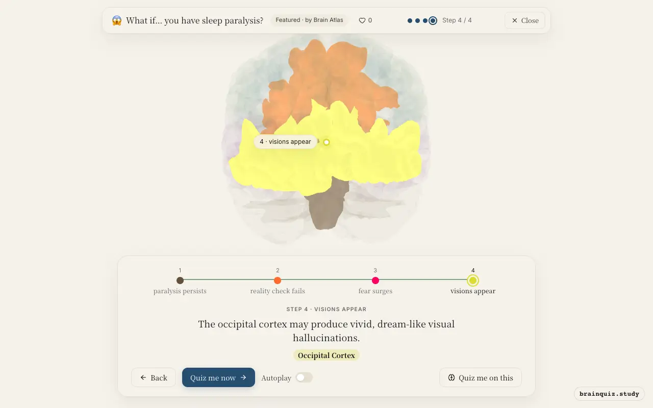
During sleep paralysis, the brainstem maintains REM atonia, causing paralysis, while the amygdala overreacts to perceived threats and the prefrontal cortex may be underactive, impairing rational thought. Visual hallucinations may arise from spontaneous occipital activity.
Watch it light up on the 3D brain
Brain regions that light up
| Order | Region | What it does here |
|---|---|---|
| 1 | Brainstem | The brainstem continues sending atonia signals to the spinal cord, paralyzing voluntary muscles. |
| 2 | Prefrontal Cortex | The prefrontal cortex may be underactive, impairing executive function and self-awareness. |
| 3 | Amygdala | The amygdala becomes highly active, generating intense fear and a sense of threat. |
| 4 | Occipital Cortex | The occipital cortex may produce vivid, dream-like visual hallucinations. |
1. Brainstem: paralysis persists
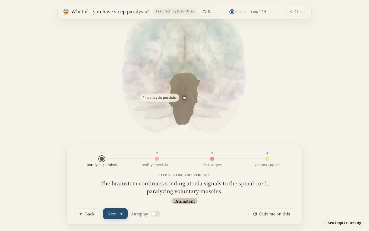
Normally, REM sleep suppresses motor neurons to prevent acting out dreams. During sleep paralysis, this atonia persists even as the cortex wakes, leaving the person unable to move or speak.
2. Prefrontal Cortex: reality check fails
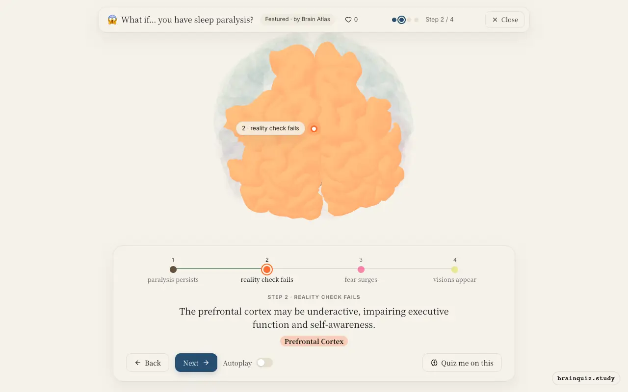
Reduced activity in the prefrontal cortex may mean the person cannot rationally evaluate the experience, so hallucinations and paralysis feel terrifyingly real. This region would normally help override fear.
3. Amygdala: fear surges
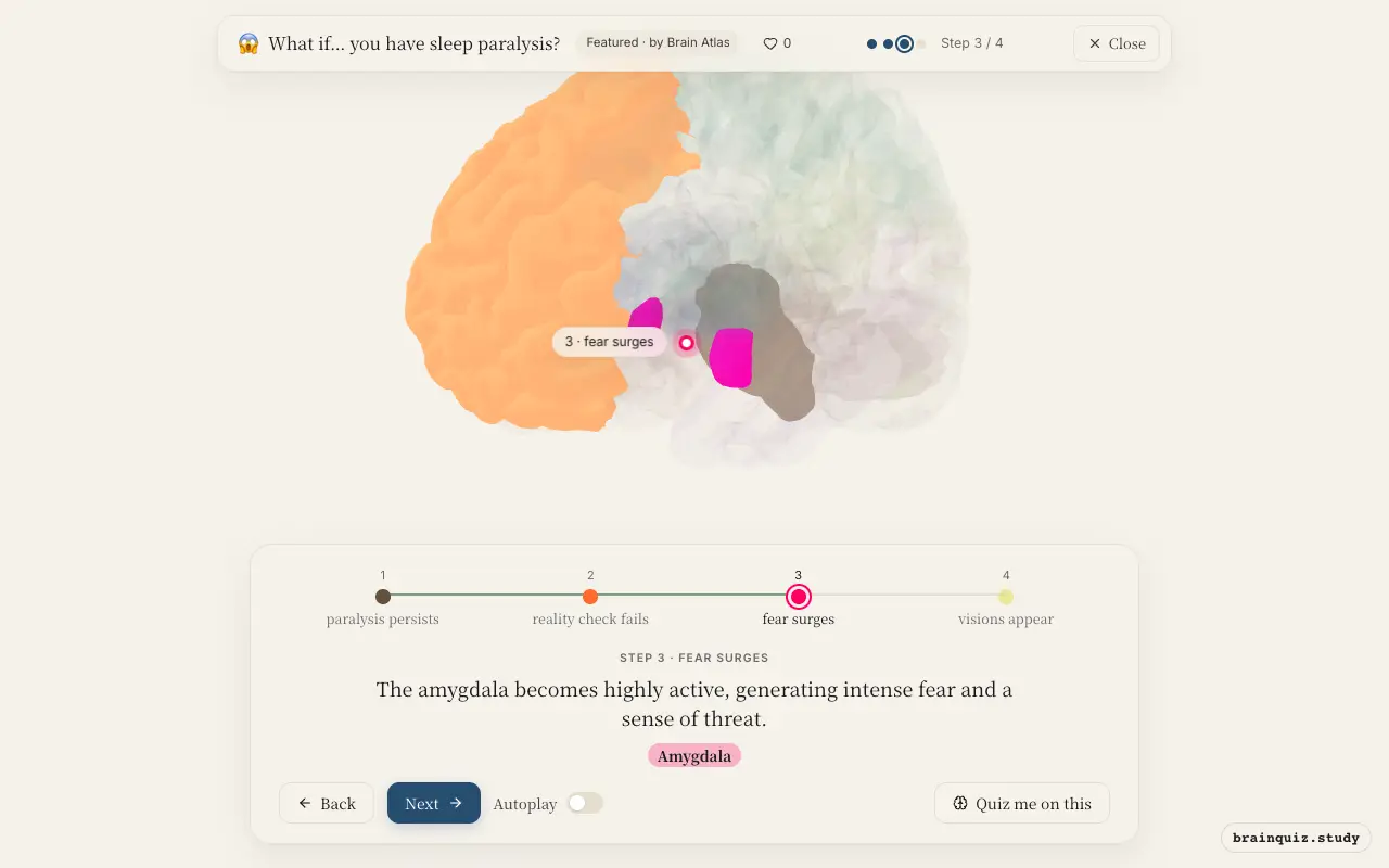
The amygdala is the brain's alarm system, and during sleep paralysis it is thought to be overactive, creating the feeling of a menacing presence or impending doom.
4. Occipital Cortex: visions appear
Without normal sensory input, the occipital cortex may fire spontaneously, generating images such as shadowy figures or intruders. These hallucinations are often consistent with the fear response.
Good to know
- Sleep paralysis occurs during transitions between REM sleep and wakefulness, and studies suggest up to 8% of the general population experiences it.
- The experience is often accompanied by hypnagogic or hypnopompic hallucinations, which are vivid dream-like perceptions.
- Sleep paralysis is more common in people with narcolepsy, but can occur in healthy individuals, especially when sleep-deprived.
Sleep Paralysis vs REM Sleep Behavior Disorder
In sleep paralysis, REM atonia persists into wakefulness, causing paralysis with conscious awareness. In REM sleep behavior disorder, the atonia fails during REM sleep, so people act out their dreams physically. Both involve brainstem dysfunction, but with opposite effects on muscle tone.
Test yourself
Which brain region is primarily responsible for the muscle paralysis in sleep paralysis?
The underactivity of which region contributes to the feeling that hallucinations are real?
What role does the amygdala play in sleep paralysis?
0 of 3 answered · 0 correct
Simplified: many regions work together at once, and this shows the main players in the order they act.