What happens in your brain when you dream?
Updated
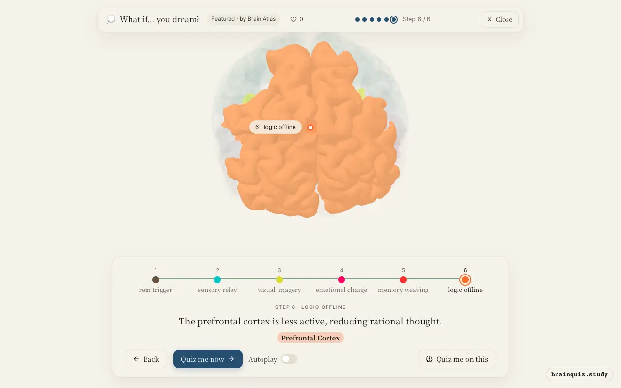
During dreams, the brainstem triggers REM sleep, the thalamus relays internal signals, the occipital cortex generates imagery, the amygdala adds emotion, and the prefrontal cortex is deactivated, allowing illogical content.
Watch it light up on the 3D brain
Brain regions that light up
| Order | Region | What it does here |
|---|---|---|
| 1 | Brainstem | The brainstem initiates REM sleep, sending signals upward. |
| 2 | Thalamus | The thalamus relays internal signals to the cortex, bypassing external input. |
| 3 | Occipital Cortex | The occipital cortex generates vivid visual scenes from internal signals. |
| 4 | Amygdala | The amygdala infuses the dream with strong emotions like fear or joy. |
| 5 | Hippocampus | The hippocampus may retrieve and bind memories into a narrative. |
| 6 | Prefrontal Cortex | The prefrontal cortex is less active, reducing rational thought. |
1. Brainstem: rem trigger
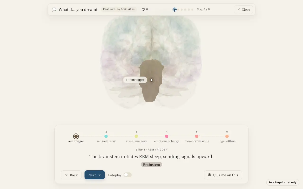
During REM sleep, brainstem nuclei become highly active and trigger the dreaming state. These nuclei send projections to the thalamus and cortex, promoting cortical arousal while simultaneously inhibiting spinal motor neurons via descending pathways. This inhibition prevents you from acting out your dreams.
2. Thalamus: sensory relay
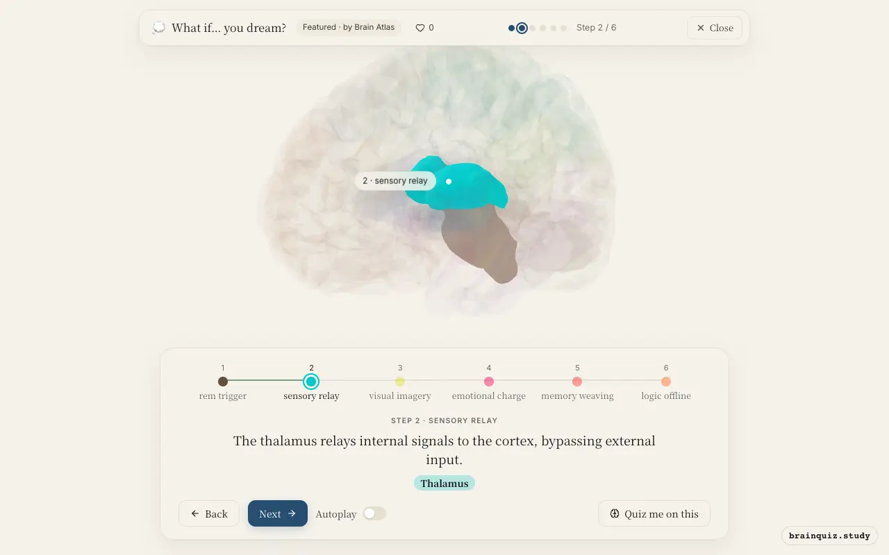
The thalamus serves as a gateway, but during REM sleep it shifts from relaying external sensory information to transmitting internally generated signals. This allows the cortex to construct dream imagery without real-world stimuli, contributing to the vivid but hallucinatory quality of dreams.
3. Occipital Cortex: visual imagery
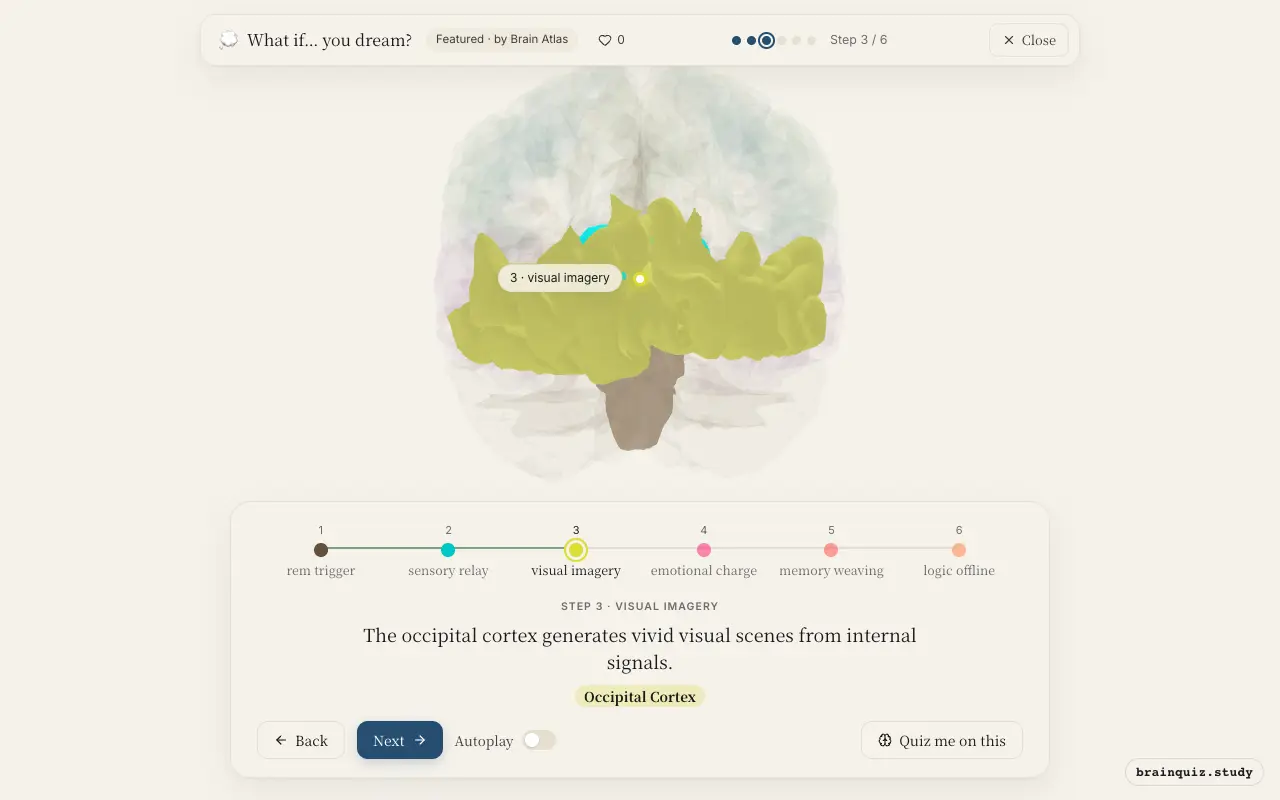
Higher-order visual association areas in the occipital cortex become active during dreaming, producing rich visual imagery. Unlike waking perception, this activity is driven top-down by memory and emotion rather than bottom-up by retinal input, which is why dream scenes can be bizarre.
4. Amygdala: emotional charge
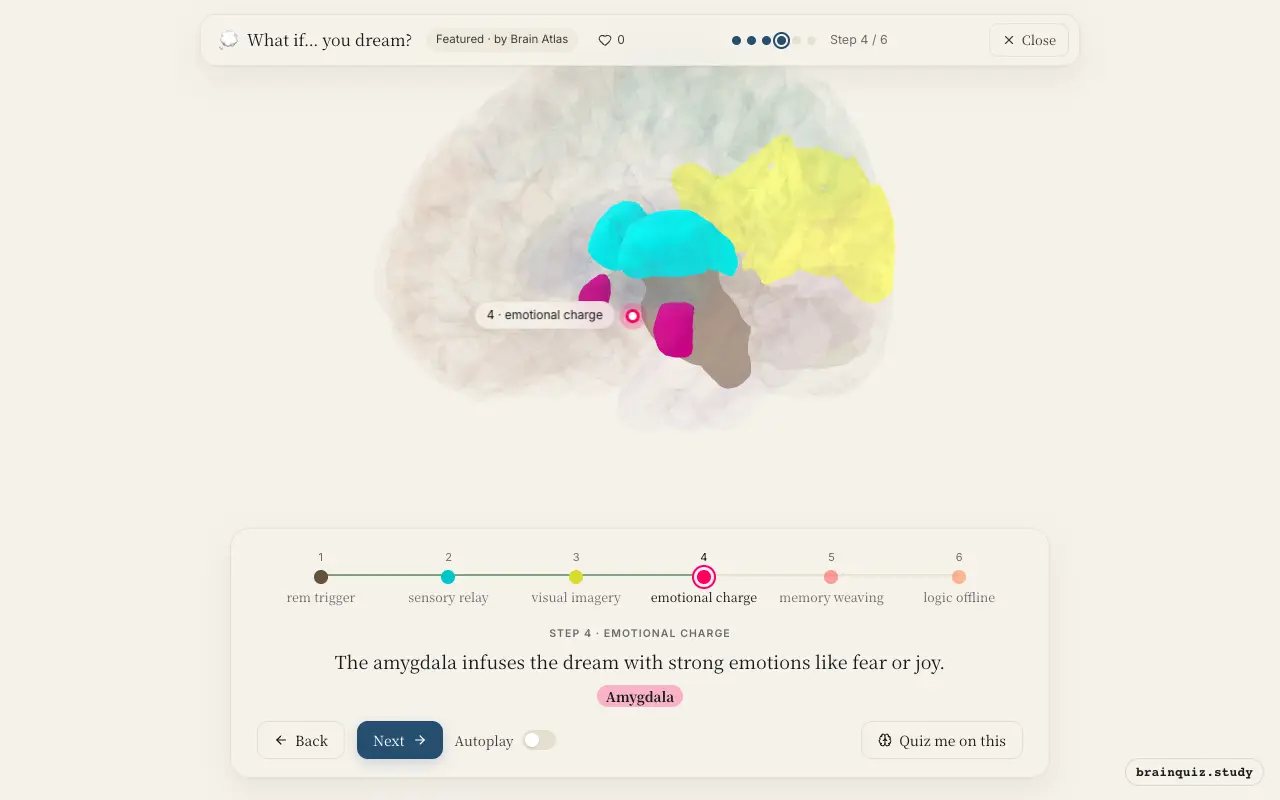
The amygdala is highly active during REM sleep, coloring dream content with intense emotions. This activity helps explain why dreams often feel emotionally vivid and why negative emotions, especially fear and anxiety, are common in nightmares. This emotional tagging may also aid memory consolidation.
5. Hippocampus: memory weaving
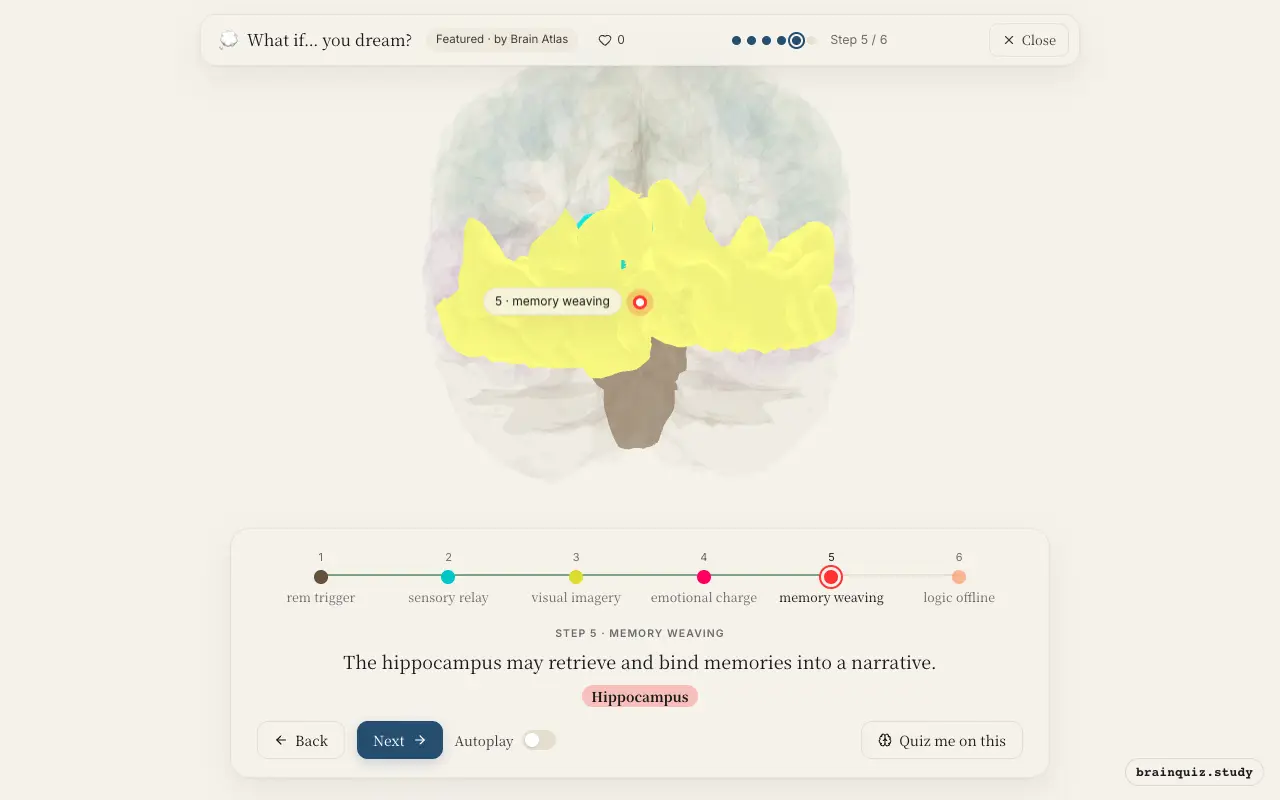
The hippocampus is thought to be active during dreaming, integrating fragments of recent and remote memories to create coherent storylines. This process may support memory consolidation, as the hippocampus replays and reorganizes experiences during sleep, helping to transfer them to long-term storage. Dreams may reflect this replay.
6. Prefrontal Cortex: logic offline
The prefrontal cortex, responsible for executive functions and reality testing, shows reduced activity during REM sleep. This deactivation explains why dreams often lack logical coherence, critical judgment, and self-awareness, allowing bizarre scenarios to unfold without question. Some lucid dreamers may show partial reactivation of this region.
Good to know
- Dreaming occurs predominantly during REM sleep, but can also happen in non-REM stages, though less vividly.
- The brainstem's inhibition of motor neurons during REM sleep causes temporary muscle paralysis, known as atonia.
- The amygdala and hippocampus show increased activity during REM sleep, while the prefrontal cortex shows decreased activity, a pattern linked to emotional memory processing.
Dreaming vs Lucid Dreaming
During normal dreaming, the prefrontal cortex is deactivated, leading to illogical scenarios. In lucid dreaming, the prefrontal cortex becomes partially reactivated, allowing the dreamer to recognize the dream state and sometimes exert control over the narrative.
Test yourself
Which brain region is most responsible for generating the visual imagery in dreams?
Why do dreams often feel illogical and bizarre?
What is the role of the amygdala during dreaming?
0 of 3 answered · 0 correct
Simplified: many regions work together at once, and this shows the main players in the order they act.