What happens in your brain when you have a nightmare?
Updated
During a nightmare, the brainstem triggers REM sleep, the amygdala fires intense fear, the hippocampus weaves memories into the narrative, the visual cortex creates vivid imagery, while the prefrontal cortex is underactive so you can't reality-check.
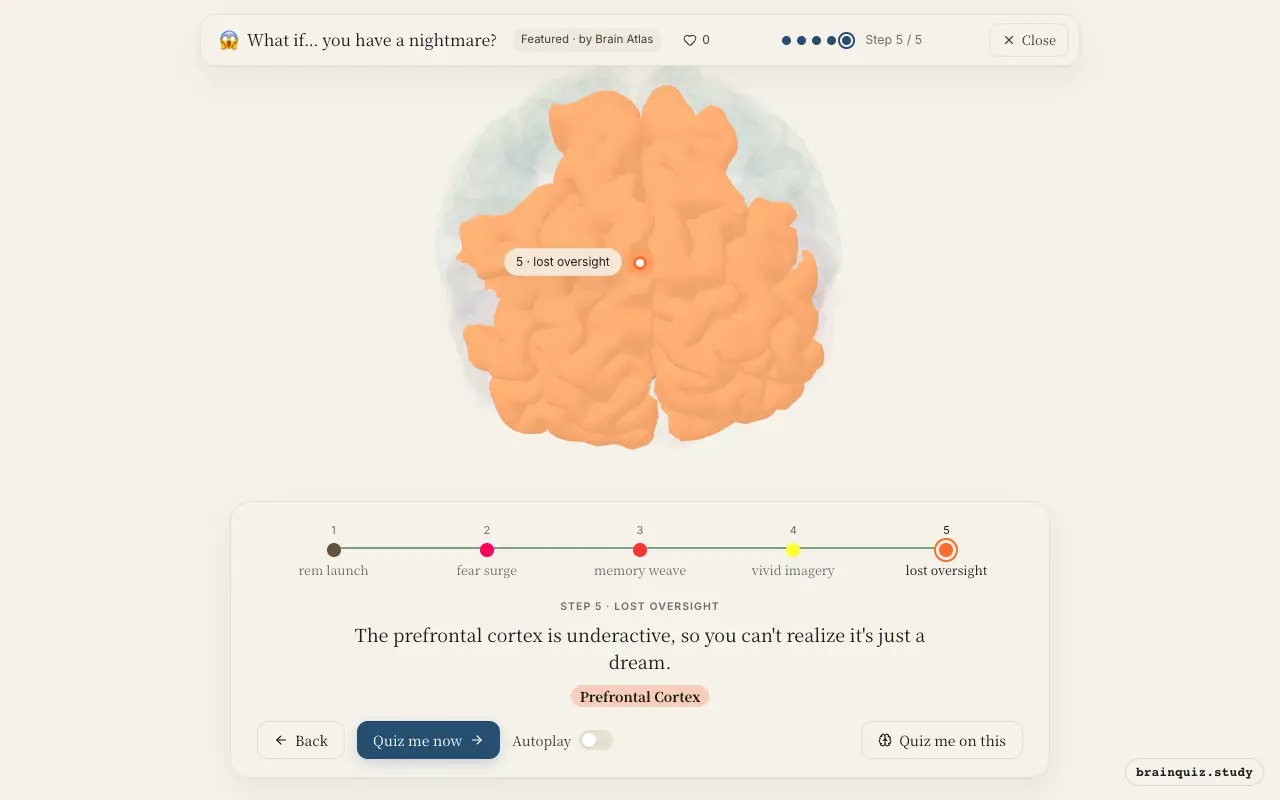
Watch it light up on the 3D brain
Brain regions that light up
| Order | Region | What it does here |
|---|---|---|
| 1 | Brainstem | The brainstem triggers REM sleep, generating the vivid dream state. |
| 2 | Amygdala | The amygdala fires intensely, producing the terror that defines a nightmare. |
| 3 | Hippocampus | The hippocampus reactivates memories, weaving them into the dream's scary narrative. |
| 4 | Visual Cortex | The visual cortex constructs the frightening images you see in the nightmare. |
| 5 | Prefrontal Cortex | The prefrontal cortex is underactive, so you can't realize it's just a dream. |
1. Brainstem: rem launch
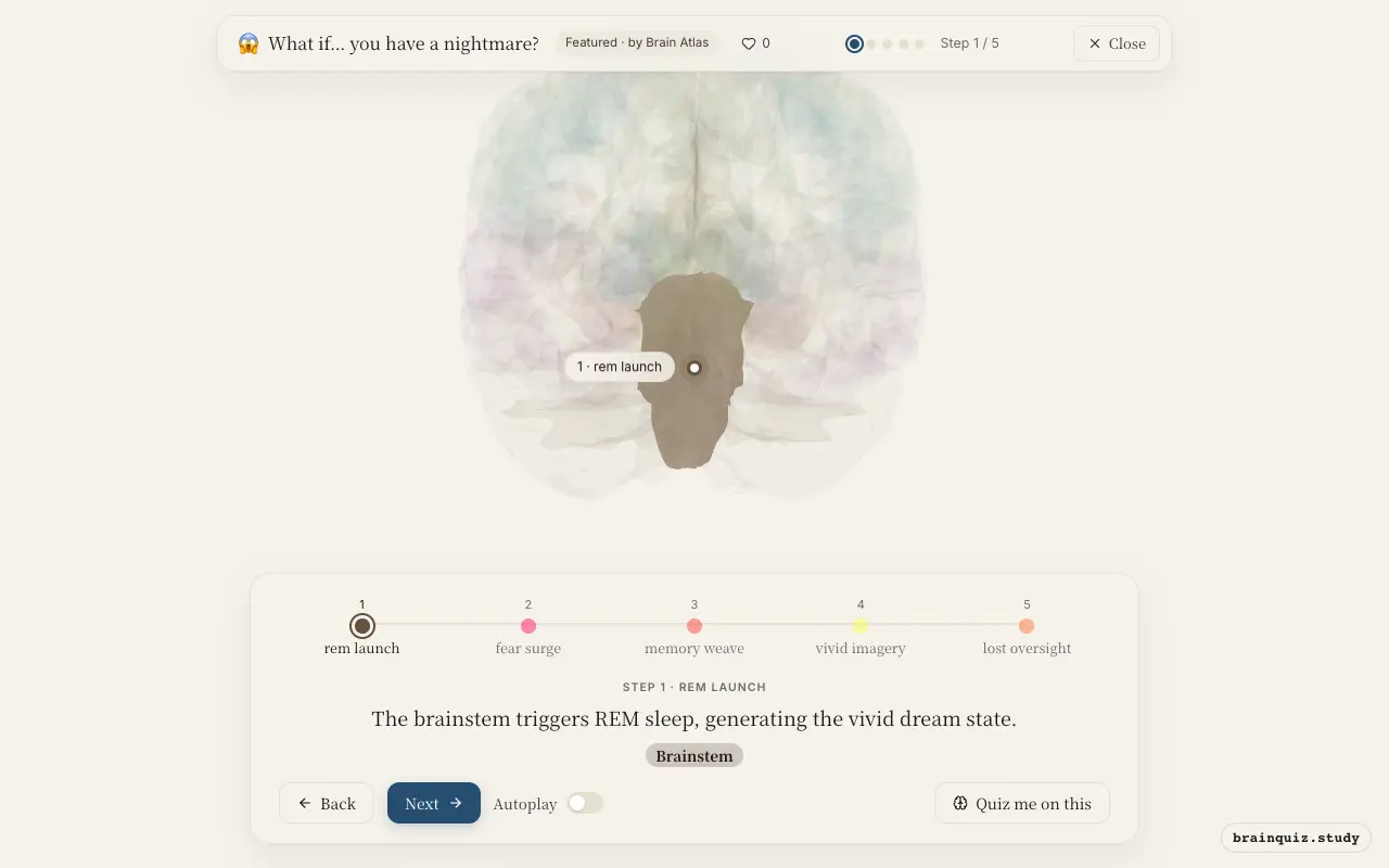
During REM sleep, brainstem nuclei such as the pedunculopontine and laterodorsal tegmental nuclei become active, causing cortical arousal and rapid eye movements. This state is permissive for dreaming, including nightmares. The brainstem also inhibits spinal motor neurons, preventing you from acting out the dream.
2. Amygdala: fear surge
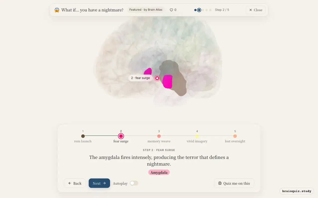
The amygdala is hyperactive during nightmares, responding as if to a real threat. It processes fear and emotional salience, and its output to the hypothalamus and brainstem triggers a stress response, even though the threat is imaginary. This activity is thought to be even stronger in people with frequent nightmares.
3. Hippocampus: memory weave
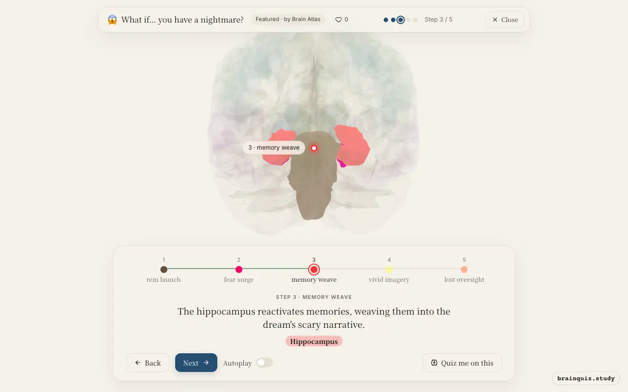
The hippocampus retrieves and integrates memory fragments, especially emotionally charged ones, into a coherent story. During sleep, it replays experiences for consolidation, but in nightmares this process may go awry, mixing fearful memories into the dream plot. This can make the nightmare feel disturbingly personal.
4. Visual Cortex: vivid imagery
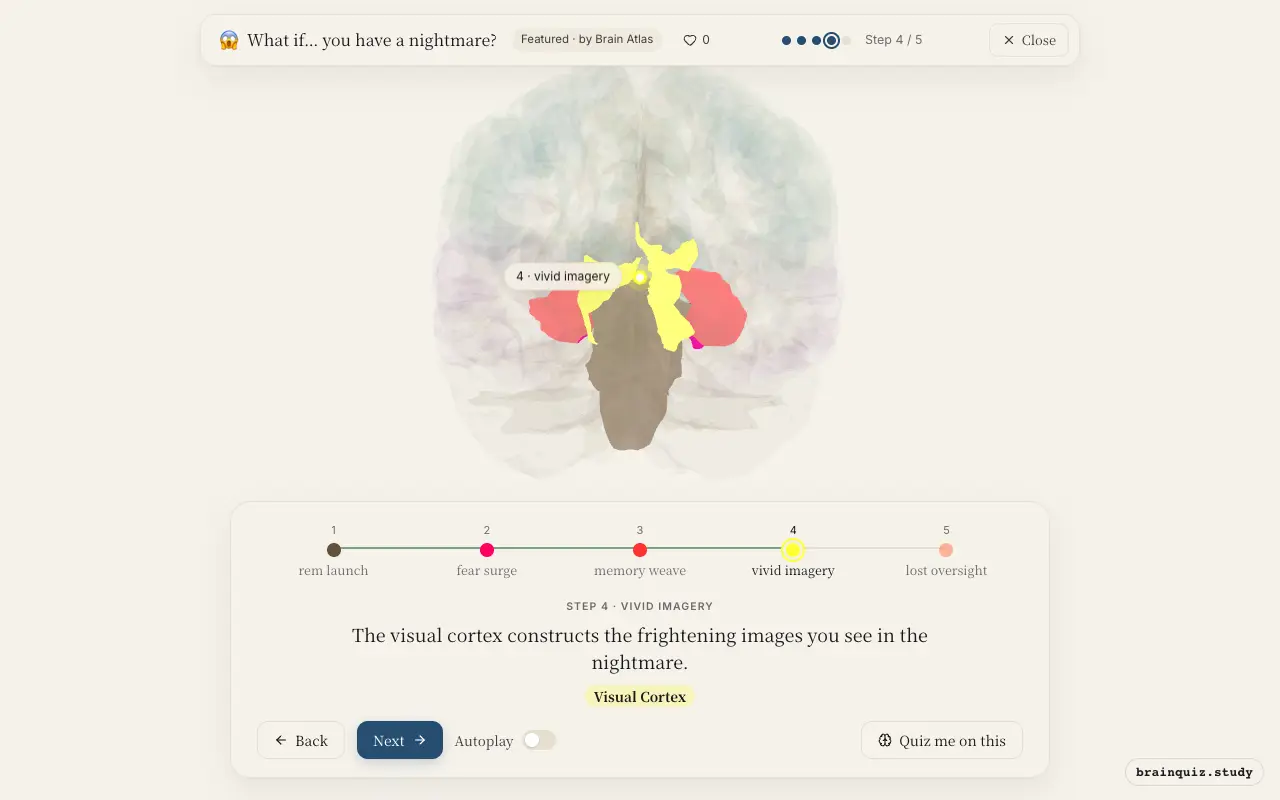
Even without external visual input, the visual cortex is active during REM sleep, generating internal imagery. In nightmares, this activity can be especially intense, producing detailed and horrifying scenes. The visual cortex is driven by feedback from the limbic system, giving the dream its cinematic quality.
5. Prefrontal Cortex: lost oversight
The dorsolateral prefrontal cortex, critical for logic and reality testing, shows reduced activity during REM sleep. This makes it hard to question the bizarre events of a nightmare. Without this top-down control, the amygdala's fear response goes unchecked, sustaining the nightmare until you wake up.
Good to know
- Nightmares occur almost exclusively during REM sleep, which is characterized by rapid eye movements and muscle atonia.
- Children experience more nightmares than adults, possibly due to higher emotional reactivity in the developing amygdala.
- The amygdala is highly active during REM sleep, which may explain why dreams are often emotionally intense.
Nightmare vs normal dream
In a normal dream, the amygdala is less active, allowing pleasant or neutral content. During a nightmare, amygdala activity spikes and prefrontal control is weaker, so fear dominates. Both occur in REM sleep, but the emotional intensity and lack of insight distinguish them.
Test yourself
Which brain region is primarily responsible for generating the fear in a nightmare?
Why can't you usually realize you're dreaming during a nightmare?
During which sleep stage do most nightmares occur?
0 of 3 answered · 0 correct
Simplified: many regions work together at once, and this shows the main players in the order they act.