Visual Cortex — Function, Location & Quiz
Primary visual cortex is the first cortical destination for visual information, arranged as a retinotopic map around the calcarine sulcus. It should not be treated as the whole occipital lobe: V1 extracts early features, while extrastriate areas specialize further in colour, motion and form.
Its orderly map makes visual-field loss anatomically precise. Damage produces a contralateral field defect, yet some patients can still respond to unseen stimuli through subcortical pathways—a dissociation known as blindsight rather than restored conscious vision.
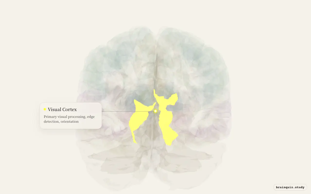
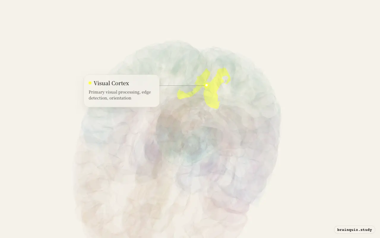
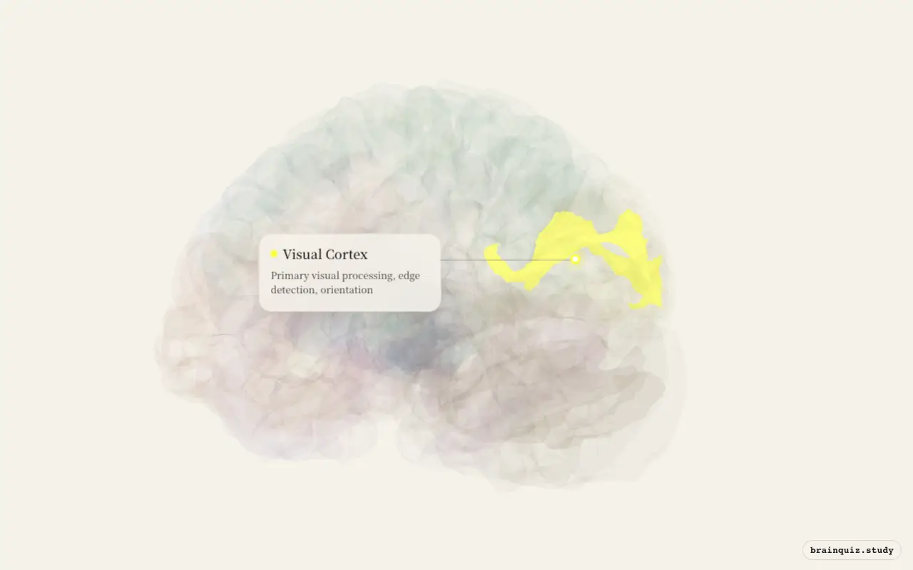
Test this anatomy in the Visual Field Defects Quiz: Localize the Lesion or explore it on the interactive 3D brain.
Where is the Visual Cortex located?
Primary visual processing, edge detection, orientation.
What does the Visual Cortex do?
- First cortical processing of visual information
- Edge detection and orientation selectivity
- Ocular dominance columns (binocular vision)
- Retinotopic organization — maps visual field
Visual Cortex pathways and connections
- LGN (lateral geniculate nucleus) → V1 (optic radiation)
- V1 → V2 → dorsal stream (V3, MT/V5 → parietal — 'where/how')
- V1 → V2 → ventral stream (V4 → IT — 'what')
What happens when the Visual Cortex is damaged?
- Lesion → contralateral homonymous hemianopia (scotoma)
- Bilateral V1 lesion → cortical blindness (Anton's syndrome if unaware)
- Blindsight — V1 damage but subcortical pathway (superior colliculus) allows unconscious vision
Visual Cortex key facts
Atlas category: cortical. Brodmann areas or landmark: BA 17 (V1 — primary visual cortex, striate cortex).
- Hubel & Wiesel (Nobel 1981): simple cells, complex cells, hypercomplex cells
- Simple cells respond to oriented edges at specific positions
- Critical period: monocular deprivation during development → permanent loss of binocularity
- Largest cortical area devoted to foveal (central) vision — cortical magnification
Visual Cortex exam tip
V1 = BA 17 = striate cortex = calcarine sulcus. Hubel & Wiesel's hierarchy: LGN → simple → complex → hypercomplex.
Common questions
Where is the primary visual cortex?
It lies around the calcarine sulcus on the medial occipital surface and corresponds to Brodmann area 17, also called striate cortex or V1.
What does V1 do?
V1 performs the first cortical analysis of visual input, representing the visual field retinotopically and responding to features such as oriented edges.
What is blindsight?
Blindsight is above-chance visual responding without conscious sight after V1 damage. Subcortical routes involving structures such as the superior colliculus can still guide limited behaviour.
When does the Visual Cortex light up?
- 🌌 When you feel awe looking at the stars: The visual cortex decodes the points of light as stars against the dark sky.
- 🍄 When you take magic mushrooms: Visual cortex becomes overactive even without external visual stimuli.
- 🤔 When you get déjà vu: The visual cortex processes the current scene, extracting basic features like edges and motion.
- 🚪 When you walk into a room and forget why: Your visual cortex rapidly processes the layout and objects of the new room.
- 📱 When you scroll social media: Your visual cortex decodes the rapidly changing images and text on the screen.
- 📱 When you get a notification on your phone: If the screen lights up, the visual cortex detects the change in your peripheral or foveal vision.
- 🚗 When you drive a familiar route on autopilot: Your visual cortex processes the familiar road, but attention is reduced.
- 🧭 When you get lost in a new city: Your visual cortex processes the streams of unfamiliar buildings, cars, and people.