What happens in your brain when you scroll social media?
Updated
Visual cortex processes the feed, amygdala flags emotionally charged posts, nucleus accumbens drives reward anticipation, prefrontal cortex decides to keep scrolling or stop.
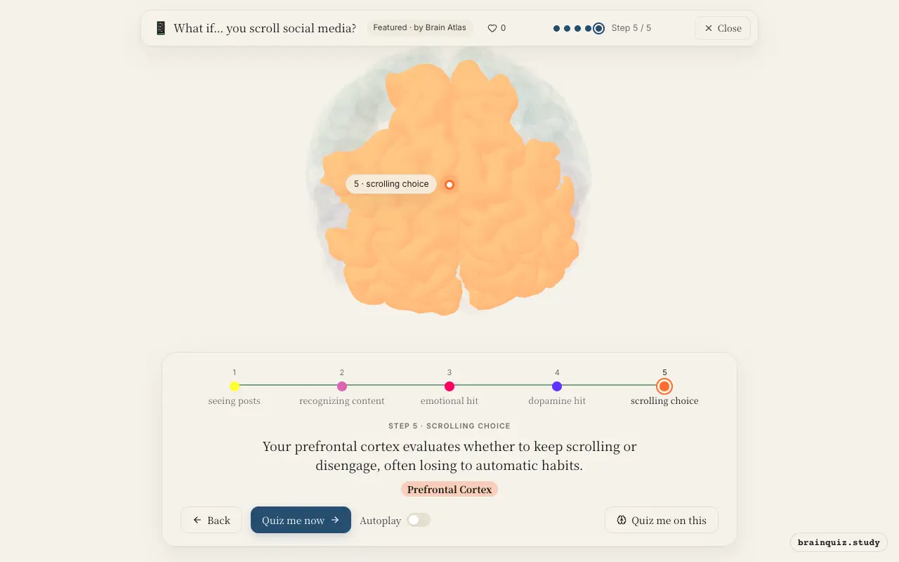
Watch it light up on the 3D brain
Brain regions that light up
| Order | Region | What it does here |
|---|---|---|
| 1 | Visual Cortex | Your visual cortex decodes the rapidly changing images and text on the screen. |
| 2 | Fusiform Gyrus | The fusiform gyrus identifies familiar faces, words, and meaningful objects in the feed. |
| 3 | Amygdala | Your amygdala tags posts with emotional weight, especially negative or arousing content. |
| 4 | Nucleus Accumbens | The nucleus accumbens signals the anticipation of reward, making you want to keep scrolling. |
| 5 | Prefrontal Cortex | Your prefrontal cortex evaluates whether to keep scrolling or disengage, often losing to automatic habits. |
1. Visual Cortex: seeing posts
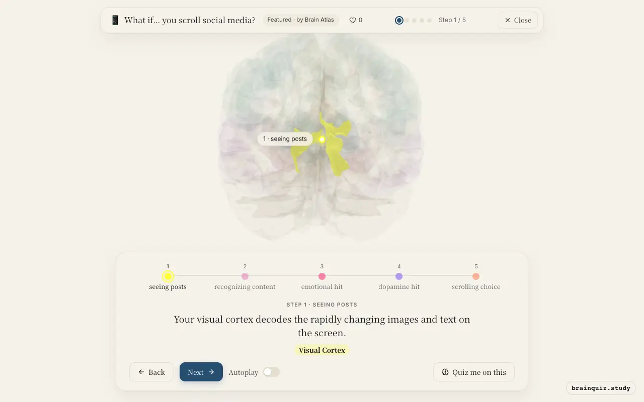
The primary visual cortex processes basic features like edges and orientation, while association areas work on motion and color. This rapid stream of visual information is the entry point for every post.
2. Fusiform Gyrus: recognizing content
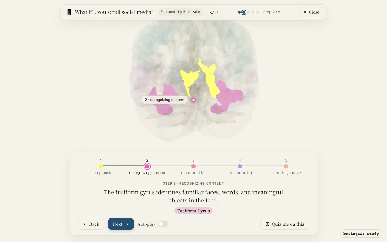
The fusiform face area and visual word form area allow quick recognition of friends' photos and readable headlines. This fast identification is crucial for determining what deserves attention.
3. Amygdala: emotional hit
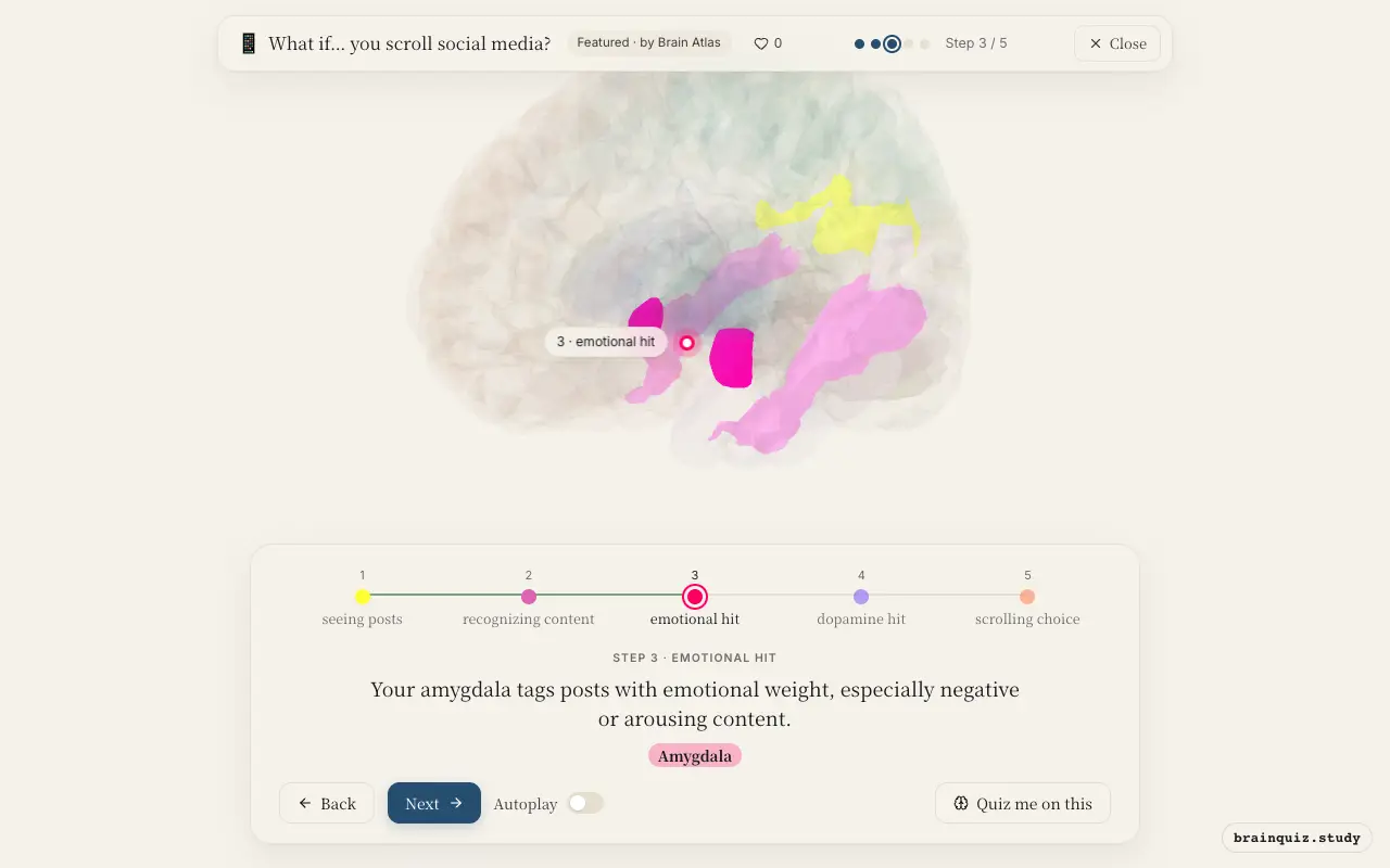
The amygdala responds rapidly to emotionally salient stimuli, particularly threats or socially relevant cues. Its activation can make certain posts feel urgent, prompting a deeper look before conscious processing.
4. Nucleus Accumbens: dopamine hit
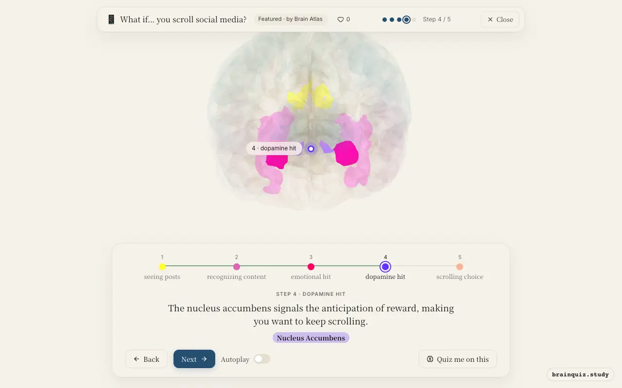
Dopamine release in the nucleus accumbens occurs not just for rewards but for the expectation of reward. Variable social feedback—likes, comments—turns scrolling into an unpredictable reward schedule, strengthening the habit.
5. Prefrontal Cortex: scrolling choice
The dorsolateral prefrontal cortex exerts top-down control, but frequent social media use may weaken its grip over habitual scrolling. Orbitofrontal cortex also weighs value, but the immediate reward pathway often overrides long-term goals.
Good to know
- Social media platforms use variable reward schedules, similar to slot machines, which rely on dopamine signaling in the nucleus accumbens.
- The default mode network, including the posterior cingulate cortex, becomes active during mind-wandering, which can occur while passively scrolling.
- Scrolling is a form of intermittent reinforcement; unpredictable rewards make the behavior resistant to extinction.
Scrolling vs reading a book
Reading a book may engage language and memory regions like Wernicke's area and hippocampus more strongly, while social media scrolling may heavily recruit reward and emotional circuits (nucleus accumbens, amygdala) with variable prefrontal engagement. The continuous novelty of a feed contrasts with the sustained attention of linear reading.
Test yourself
Which region is most responsible for the 'wanting' to keep scrolling?
What role does the amygdala play when you see an emotionally charged post?
Why does scrolling become habit-forming?
0 of 3 answered · 0 correct
Simplified: many regions work together at once, and this shows the main players in the order they act.