What Is the Visual Network? Brain Regions and Function
Updated
The Visual Network is the brain system that processes visual information. Core regions include the occipital cortex, fusiform gyrus, and posterior parietal cortex. It handles object recognition and spatial vision.
The visual system was mapped in the late 20th century through lesion studies and neuroimaging. Ungerleider and Mishkin's two-stream model from 1982 shaped understanding of object and spatial vision. This network matters because it underpins how we see, recognize, and interact with the world, and its breakdown causes specific visual disorders.
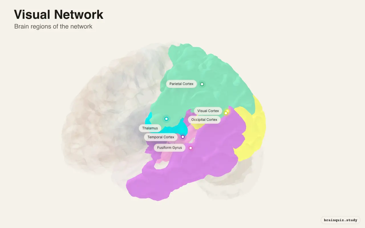
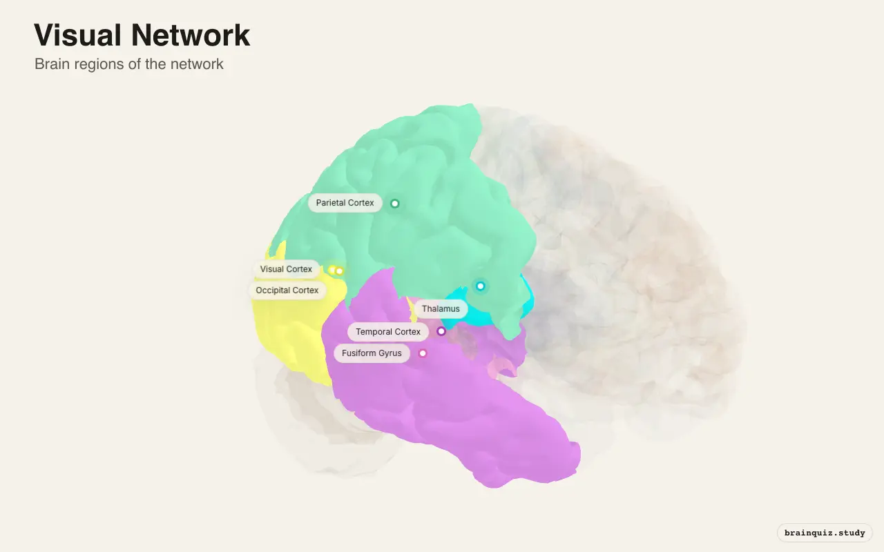
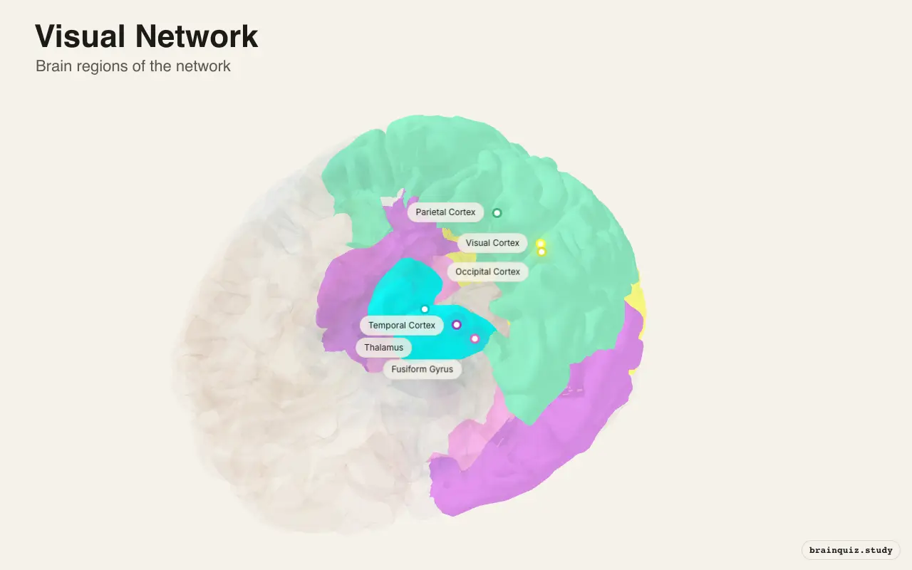
Which brain regions are in the Visual Network?
- Visual Cortex: Primary visual cortex (V1) extracts local features such as edges, orientation, and spatial frequency, sending signals to higher areas.
- Occipital Cortex: Extrastriate occipital regions (V2–V5/MT) process color, form, and motion, feeding both ventral and dorsal streams.
- Fusiform Gyrus: The fusiform gyrus, particularly the fusiform face area (FFA), performs high-level object and face recognition.
- Parietal Cortex: Posterior parietal cortex integrates spatial information for visually guided reaching and spatial attention.
- Temporal Cortex: Inferotemporal cortex (IT) represents complex object shapes and supports semantic visual memory.
- Thalamus: The lateral geniculate nucleus of the thalamus relays retinal signals to V1 and modulates visual attention.
What does the Visual Network do?
What does the Visual Network do? The Visual Network is the set of interconnected brain regions that transforms retinal input into coherent visual percepts. It extracts basic features in V1, then splits into two streams: the ventral stream identifies objects and faces, while the dorsal stream locates objects and guides actions. The network also processes motion, color, and depth, enabling us to navigate and interact with our environment.
- Edge and orientation detection — V1 neurons respond to edges, orientations, and spatial frequencies, forming the first cortical representation of shape.
- Color and form processing — Areas V4 and the fusiform gyrus analyze color and complex form, contributing to object constancy.
- Motion and optic flow — Area V5/MT detects visual motion and optic flow, critical for perceiving self-motion and moving objects.
- Face and object recognition — The fusiform face area and nearby temporal cortex identify faces and many categories of familiar objects.
- Spatial vision and action — The dorsal stream, including posterior parietal cortex, maps spatial locations and guides reaching and grasping.
When is the Visual Network active?
The Visual Network is highly active whenever the eyes are open and visual scenes reach the retina. It responds strongly during tasks requiring detailed inspection, such as reading, recognizing faces, or judging distances. Activity rises during visual attention and falls when the eyes are closed, in darkness, or when attention is diverted to nonvisual tasks or internal thought.
- Spotting a friend's face in a crowd.
- Catching a ball thrown toward you.
- Reading text on a screen.
- Noticing a car moving in your peripheral vision.
- Reaching for a coffee mug without looking.
Visual Network vs Dorsal Attention Network
The Visual Network processes visual information, while the Dorsal Attention Network selects where and what to attend. The VN is driven primarily by bottom-up sensory input, whereas the DAN generates top-down attentional signals. The DAN biases visual cortex toward task-relevant locations or features, enhancing VN responses there and suppressing elsewhere. Together they enable goal-directed visual search and flexible prioritization of visual information.
VN vs DAN at a glance
| Aspect | Visual Network | Dorsal Attention Network |
|---|---|---|
| Primary function | Processes visual features and builds visual percepts | Allocates top-down attention to spatial locations and features |
| Signal origin | Bottom-up sensory input from retina | Endogenous goals and expectations |
| Attentional role | Receives attentional modulation that sharpens processing | Generates attentional control signals that bias sensory cortex |
| Visual specificity | Highly specialized for vision only | Modality-general; can also bias auditory or tactile processing |
| Typical activation | Active during passive viewing and visual tasks | Active during voluntary attention shifts even without visual stimuli |
What happens when the Visual Network is disrupted?
Damage or degeneration within the Visual Network can produce selective visual deficits depending on location. Lesions to primary visual cortex cause blindness in part of the visual field, while damage to higher areas can impair specific abilities like face recognition or motion perception. Some disorders emerge gradually in neurodegenerative disease, often beginning with subtle visual complaints.
- Homonymous hemianopia — Damage to one occipital lobe or its inputs causes loss of vision in the opposite half of the visual field in both eyes.
- Prosopagnosia — Injury to the fusiform face area is associated with an inability to recognize familiar faces, despite intact basic vision.
- Akinetopsia — Bilateral damage to area V5/MT can produce motion blindness, where moving objects appear as static snapshots.
- Cortical blindness — Bilateral occipital lesions may cause total vision loss; if the person denies blindness, it is called Anton syndrome.
- Posterior cortical atrophy — This visual variant of Alzheimer's disease gradually disrupts higher visual processing, leading to reading and object recognition difficulties.
How does the Visual Network work with other networks?
The Visual Network works most closely with the Dorsal Attention Network, which directs visual processing toward relevant stimuli. It also interacts with the Ventral Attention Network to reorient attention to salient or unexpected visual events. The frontoparietal control network helps maintain task goals that shape visual analysis, while the default mode network may reduce activity when the visual network is engaged in external tasks. In resting-state studies, visual regions show strong internal connectivity, often forming a distinct intrinsic network.
How do scientists find the Visual Network?
Researchers commonly identify the Visual Network using resting-state fMRI, where low-frequency fluctuations in blood oxygen level-dependent (BOLD) signal show synchronized activity among visual areas. Seed-based analysis places a seed in primary visual cortex or extrastriate cortex, while independent component analysis (ICA) separates the network automatically. A limitation is that fMRI measures hemodynamic changes indirectly, with coarse spatial and temporal resolution compared to underlying neural activity.
Key facts about the Visual Network
- The visual system's two-stream organization was proposed by Ungerleider and Mishkin in 1982, based on lesion studies in monkeys.
- Primary visual cortex (V1) has a retinotopic map, with adjacent points in visual space represented by adjacent neurons.
- The lateral geniculate nucleus of the thalamus is the main relay from the retina to V1, with six layers in primates.
Sources
- Goodale MA, Milner AD (1992). Separate visual pathways for perception and action. Trends in Neurosciences, 15(1), 20–25.
- Yeo BTT et al. (2011). The organization of the human cerebral cortex estimated by intrinsic functional connectivity. Journal of Neurophysiology, 106(3), 1125–1165.