What happens in your brain when you take an antidepressant?
Updated
Antidepressants boost serotonin and norepinephrine in the dorsal raphe and locus coeruleus, dampen amygdala overreactivity, strengthen prefrontal cortex control, and promote hippocampal neurogenesis.
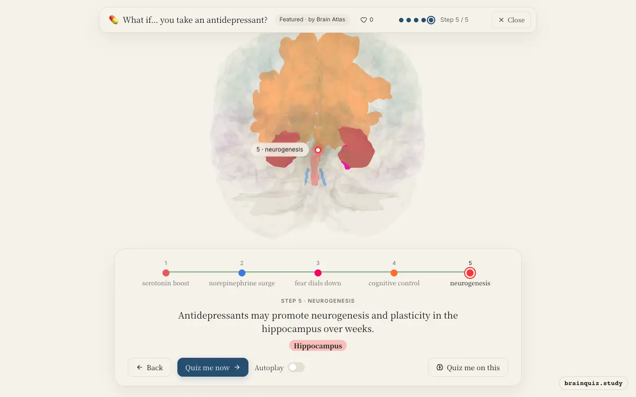
Watch it light up on the 3D brain
Brain regions that light up
| Order | Region | What it does here |
|---|---|---|
| 1 | Dorsal Raphe Nucleus | The antidepressant blocks serotonin reuptake, increasing serotonin in the dorsal raphe. |
| 2 | Locus Coeruleus | Some antidepressants also block norepinephrine reuptake, raising levels in the locus coeruleus. |
| 3 | Amygdala | Elevated monoamines reduce the amygdala's exaggerated response to negative emotional stimuli. |
| 4 | Prefrontal Cortex | The prefrontal cortex gains stronger top-down regulation over emotional areas. |
| 5 | Hippocampus | Antidepressants may promote neurogenesis and plasticity in the hippocampus over weeks. |
1. Dorsal Raphe Nucleus: serotonin boost
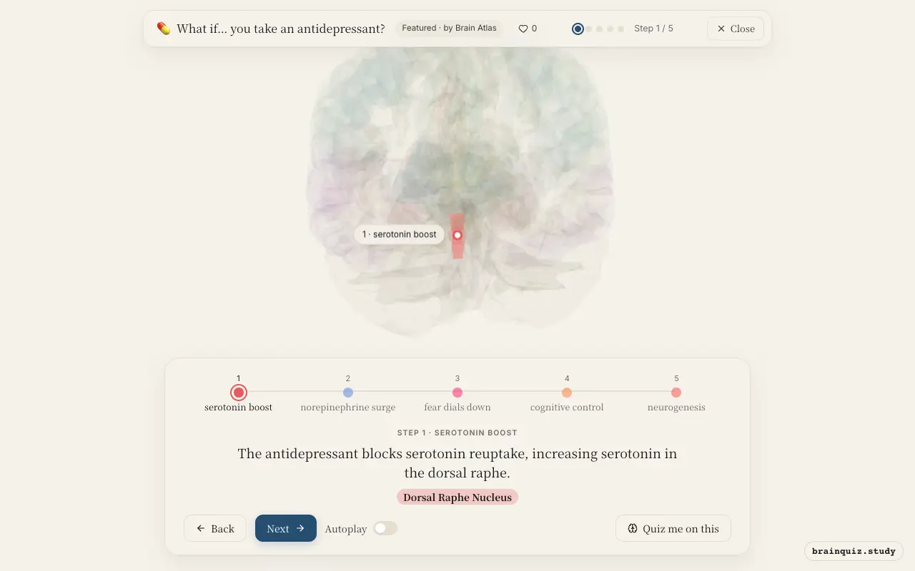
The dorsal raphe nucleus is the main source of serotonin in the brain, projecting to cortex and limbic areas. Antidepressants like SSRIs block the serotonin transporter, initially increasing serotonin at cell bodies. Over weeks, autoreceptors desensitize, enhancing serotonergic signaling throughout the brain.
2. Locus Coeruleus: norepinephrine surge
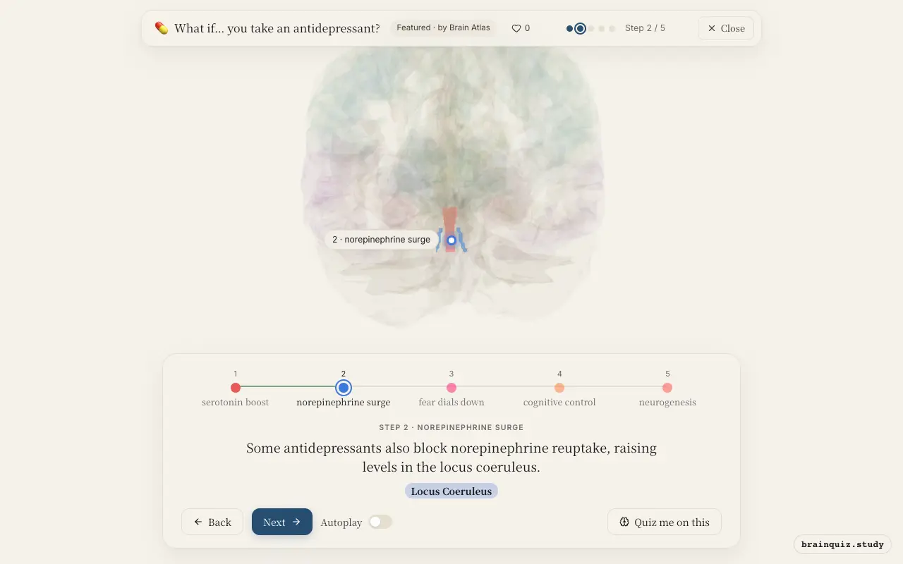
The locus coeruleus is the principal noradrenergic nucleus, involved in arousal and stress response. SNRIs or tricyclics increase norepinephrine here, contributing to improved energy, attention, and mood. Elevated norepinephrine enhances signaling to prefrontal cortex and limbic regions.
3. Amygdala: fear dials down
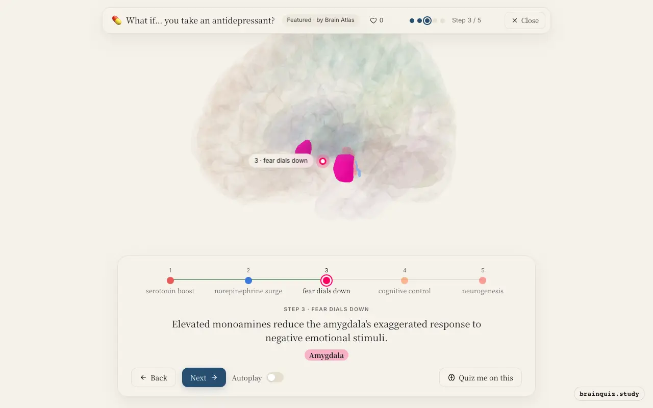
The amygdala is hyperactive in depression, leading to excessive fear and negative bias. Antidepressants normalize its activity, reducing the emotional salience of negative events. This effect is thought to occur early in treatment, even before mood lifts.
4. Prefrontal Cortex: cognitive control
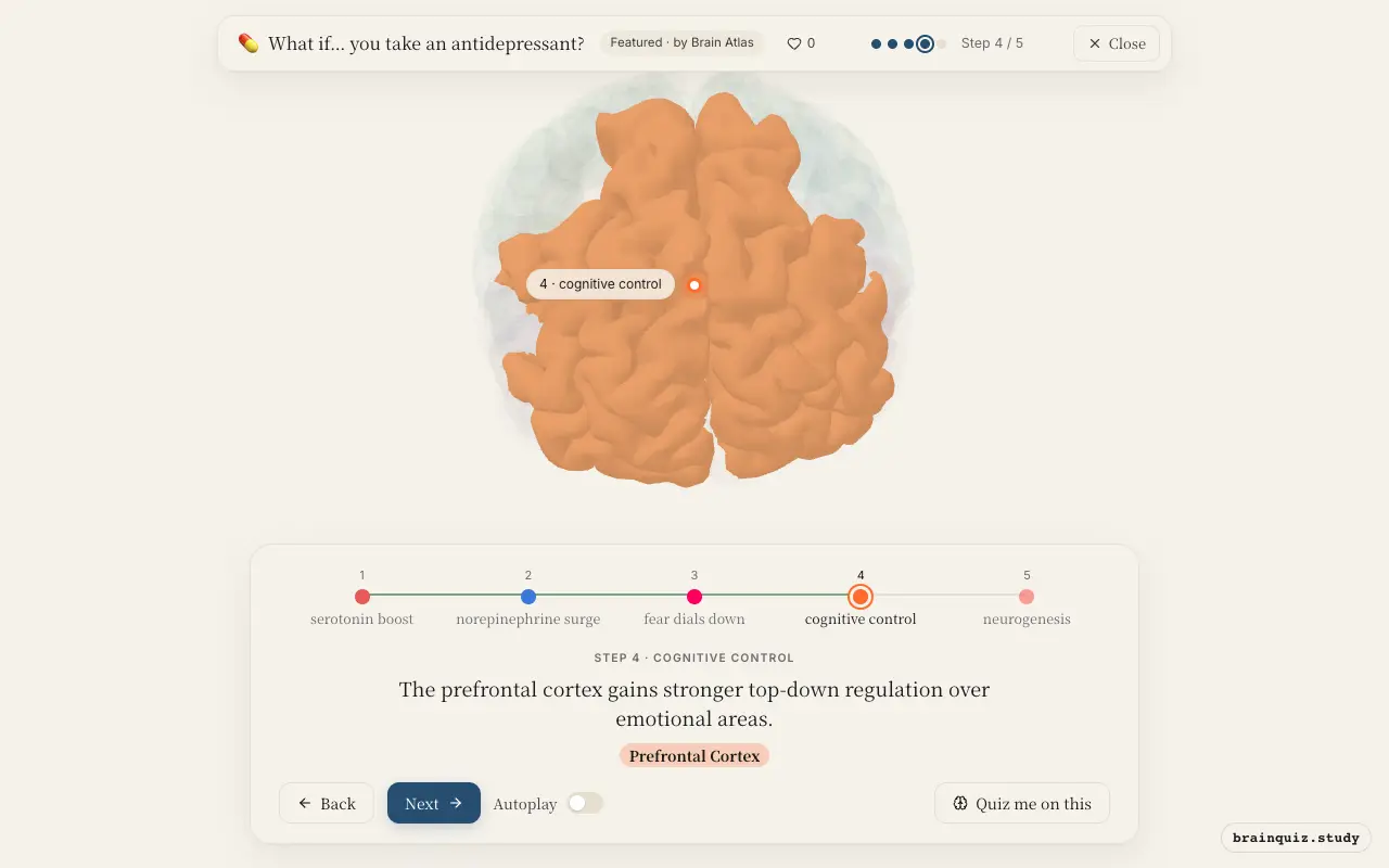
The prefrontal cortex is critical for executive function and emotion regulation. Increased serotonin and norepinephrine enhance its activity, allowing better inhibition of amygdala-driven responses. This improved cognitive control helps patients reappraise negative thoughts.
5. Hippocampus: neurogenesis
The hippocampus often shrinks in depression due to chronic stress. Antidepressants increase brain-derived neurotrophic factor and stimulate new neuron growth. This delayed neuroplasticity is thought to underlie the therapeutic lag of several weeks.
Good to know
- Most antidepressants target the serotonin transporter (SERT) to increase synaptic serotonin availability.
- The delayed therapeutic effect is thought to involve neuroplastic changes rather than immediate neurotransmitter increases.
- Different antidepressant classes (SSRI, SNRI, TCA) affect different monoamine systems.
SSRIs vs SNRIs: brain effects
SSRIs primarily increase serotonin, mainly affecting the dorsal raphe and limbic areas. SNRIs also boost norepinephrine, more strongly engaging the locus coeruleus and arousal pathways. This dual action may explain why some patients respond better to SNRIs for fatigue or pain.
Test yourself
Which brain region is the main source of serotonin and a primary site of action for SSRIs?
What long-term brain change is thought to underlie antidepressant efficacy?
How do antidepressants affect the amygdala?
0 of 3 answered · 0 correct
Simplified: many regions work together at once, and this shows the main players in the order they act. For learning only, not medical advice.