What happens in your brain when you eat until you're full?
Updated
As you eat, the brainstem relays fullness signals to the hypothalamus, which reduces hunger; the insula creates conscious fullness, the orbitofrontal cortex lowers food's reward value, and the prefrontal cortex decides to stop.
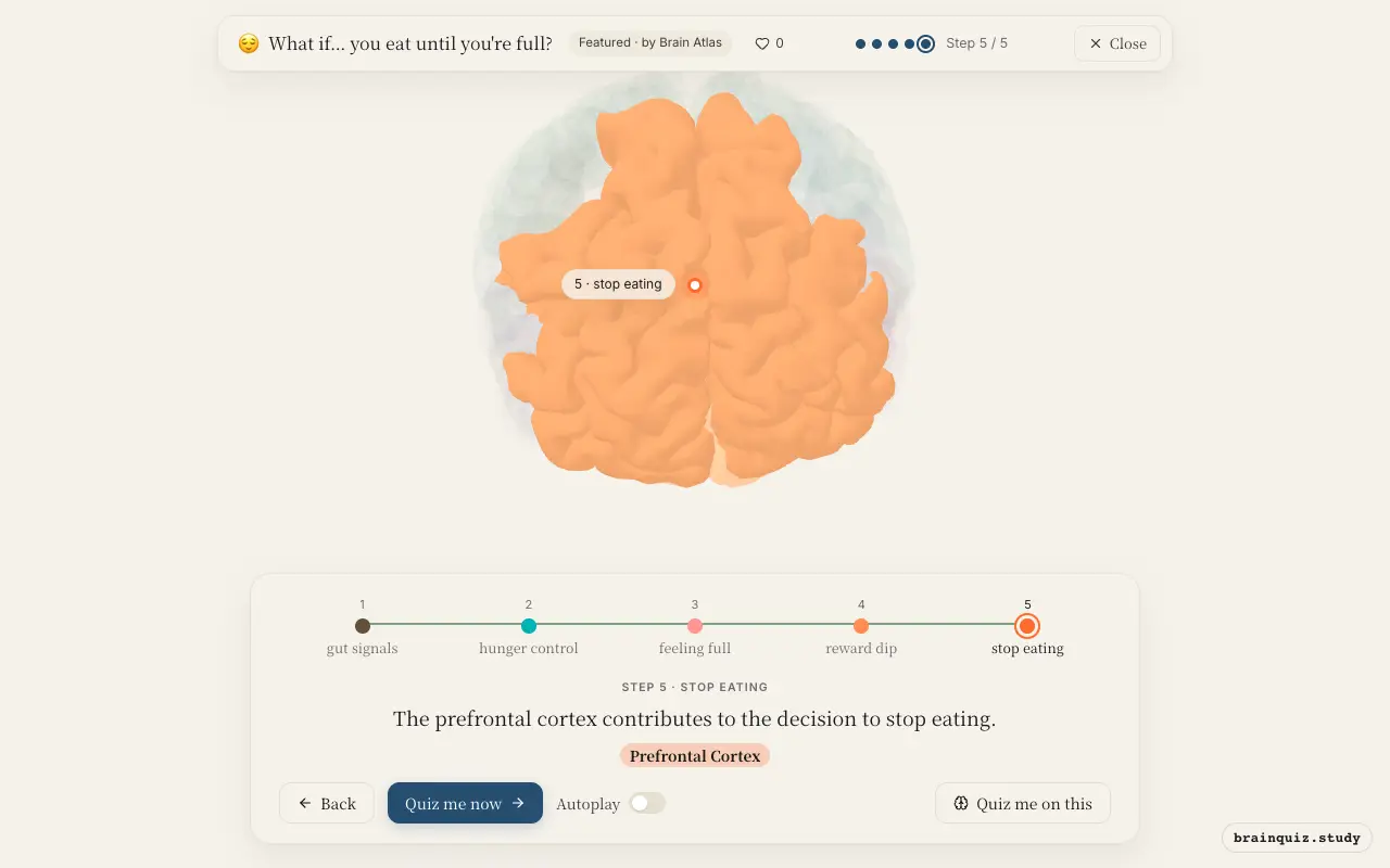
Watch it light up on the 3D brain
Brain regions that light up
| Order | Region | What it does here |
|---|---|---|
| 1 | Brainstem | Stretch receptors in your stomach send signals via the vagus nerve to the brainstem. |
| 2 | Hypothalamus | The hypothalamus integrates these signals and reduces hunger drive. |
| 3 | Insula | The insula generates the conscious feeling of fullness and discomfort. |
| 4 | Orbitofrontal Cortex | The orbitofrontal cortex decreases the reward value of food as you approach fullness. |
| 5 | Prefrontal Cortex | The prefrontal cortex contributes to the decision to stop eating. |
1. Brainstem: gut signals
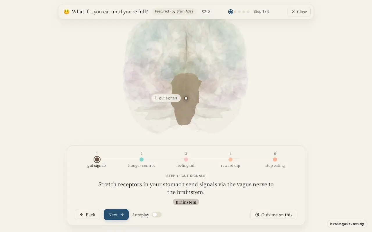
As food fills the stomach, mechanoreceptors detect distension and fire along the vagus nerve. The brainstem's nucleus of the solitary tract receives these visceral signals and relays them upward. This is the first neural step in satiety, before conscious awareness.
2. Hypothalamus: hunger control
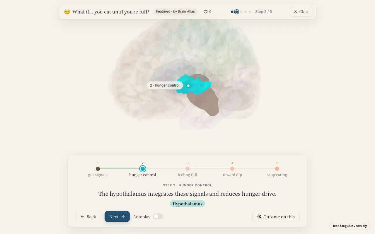
The arcuate nucleus of the hypothalamus receives input about nutrients and hormones like leptin and insulin. It then inhibits orexigenic (hunger-promoting) neurons and activates anorexigenic (satiety-promoting) neurons. This shifts the brain from seeking food to feeling satisfied, a central hub for energy balance.
3. Insula: feeling full
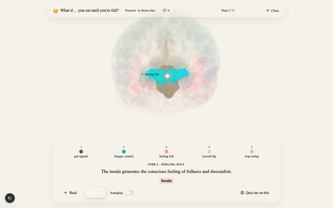
The insula processes interoceptive signals from the body, including stomach distension and satiety hormones, creating the subjective experience of fullness. It is part of a network that monitors internal state. This awareness is crucial for deciding when to stop eating.
4. Orbitofrontal Cortex: reward dip
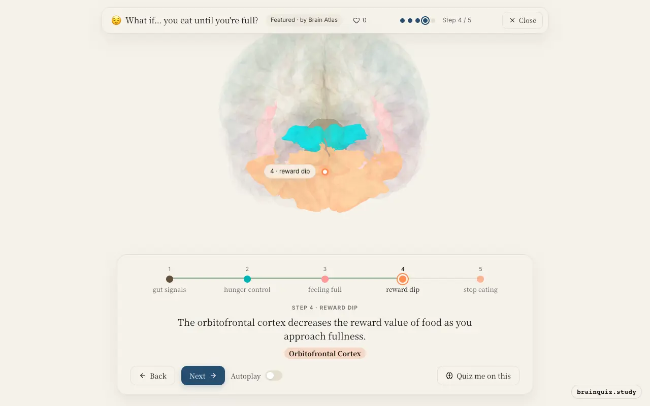
The orbitofrontal cortex encodes the current value of food based on sensory input and internal state. When full, its response to food cues diminishes markedly, making further eating far less appealing. This is a key mechanism of sensory-specific satiety, reducing desire.
5. Prefrontal Cortex: stop eating
Executive control regions in the prefrontal cortex integrate signals from the insula and orbitofrontal cortex. They influence the decision to stop eating by integrating signals from the insula and orbitofrontal cortex. This top-down control is essential for self-regulation, often preventing overeating.
Good to know
- Leptin, released from fat stores, provides a long-term signal of energy reserves to the hypothalamus.
- The vagus nerve is a major pathway for gut-brain communication during meals.
- Dopamine release in reward areas drops as satiety sets in, reducing food motivation.
Fullness vs. hunger: flip of the switch
When hungry, the hypothalamus activates hunger neurons and the orbitofrontal cortex assigns high reward value to food. As you eat and become full, these patterns reverse: the hypothalamus promotes satiety and the orbitofrontal cortex devalues food. The insula shifts from craving to discomfort.
Test yourself
Which region first receives fullness signals from the stomach?
What happens in the orbitofrontal cortex as you become full?
The insula is primarily responsible for:
0 of 3 answered · 0 correct
Simplified: many regions work together at once, and this shows the main players in the order they act.