What happens in your brain when you try to remember a face?
Updated
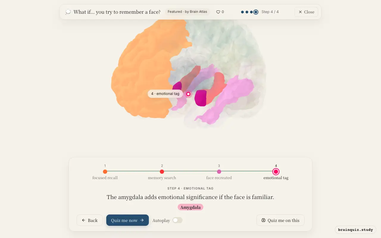
Your prefrontal cortex focuses attention, the hippocampus retrieves the memory, the fusiform gyrus reconstructs the facial image, and the amygdala adds emotional valence.
Watch it light up on the 3D brain
Brain regions that light up
| Order | Region | What it does here |
|---|---|---|
| 1 | Prefrontal Cortex | Your prefrontal cortex sets the goal and holds the face in mind. |
| 2 | Hippocampus | The hippocampus activates stored memory traces linked to the face. |
| 3 | Fusiform Gyrus | The fusiform gyrus rebuilds the facial features into a coherent image. |
| 4 | Amygdala | The amygdala adds emotional significance if the face is familiar. |
1. Prefrontal Cortex: focused recall
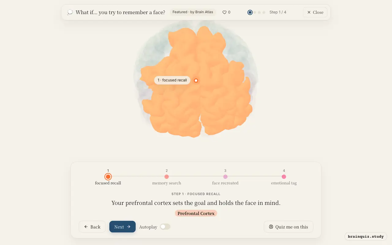
The prefrontal cortex, particularly the dorsolateral region, initiates top-down control to focus attention on the memory task. It maintains the search criteria in working memory, directing other brain regions to retrieve the relevant face. Without this executive function, recall would be haphazard.
2. Hippocampus: memory search
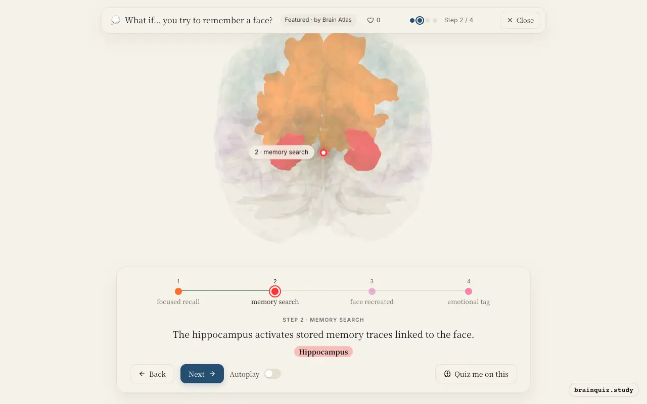
The hippocampus acts as a convergence zone, reactivating distributed cortical patterns that encode the face. Through pattern completion, it retrieves the associative network, including the person’s name and context. This process is essential for conscious recollection rather than mere familiarity.
3. Fusiform Gyrus: face recreated
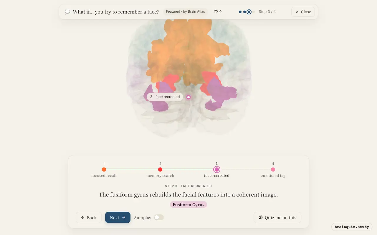
Located in the ventral temporal cortex, the fusiform face area specializes in processing facial identity. When activated during recall, it supports the vivid mental imagery of the face, allowing you to 'see' it in your mind’s eye. Damage here can cause prosopagnosia, the inability to recognize faces.
4. Amygdala: emotional tag
If the remembered face has personal or emotional relevance, the amygdala modulates the recall by tagging the memory with affective tone. This can enhance memory vividness and retrieval speed, as emotionally salient information is prioritized. However, neutral faces can activate the amygdala subconsciously.
Good to know
- The fusiform gyrus shows increased activity when viewing or imagining faces compared to other objects.
- Patients with hippocampal damage often struggle to recall detailed facial images despite intact recognition.
- Emotional faces are remembered better due to amygdala-hippocampus interactions.
Recall vs. Recognition
In recall, the prefrontal cortex and hippocampus drive the process, reconstructing the face from memory. In recognition, the fusiform gyrus signals familiarity more automatically, with less prefrontal effort. Recognition relies on a feeling of knowing, while recall requires active search.
Test yourself
Which brain region is critical for reconstructing the visual image of a face during recall?
What role does the hippocampus play when you try to remember a face?
Damage to which area might specifically impair your ability to recognize faces, even if you can see them?
0 of 3 answered · 0 correct
Simplified: many regions work together at once, and this shows the main players in the order they act.