What happens in your brain when you study with flashcards?
Updated
Seeing a flashcard activates visual cortex and fusiform gyrus for word recognition, hippocampus retrieves the answer, prefrontal cortex holds it in working memory, and anterior cingulate monitors for errors; correct answers trigger dopamine in nucleus accumbens.
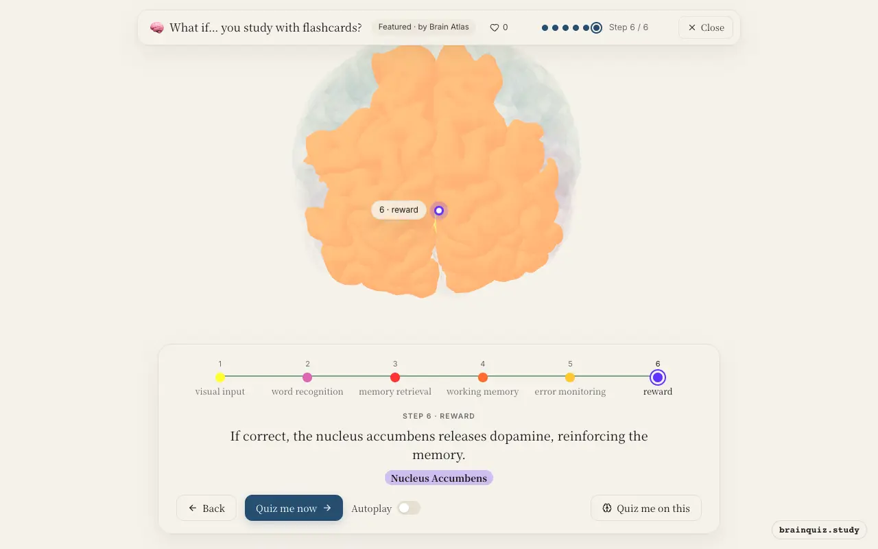
Watch it light up on the 3D brain
Brain regions that light up
| Order | Region | What it does here |
|---|---|---|
| 1 | Visual Cortex | The visual cortex decodes the letters and shapes on the flashcard. |
| 2 | Fusiform Gyrus | The fusiform gyrus recognizes the written word as a familiar visual form. |
| 3 | Hippocampus | The hippocampus retrieves the associated memory, linking the word to its meaning. |
| 4 | Prefrontal Cortex | The prefrontal cortex holds the retrieved answer in working memory while you decide to respond. |
| 5 | Anterior Cingulate Cortex | The anterior cingulate cortex compares your answer to the expected one, detecting any mismatch. |
| 6 | Nucleus Accumbens | If correct, the nucleus accumbens releases dopamine, reinforcing the memory. |
1. Visual Cortex: visual input
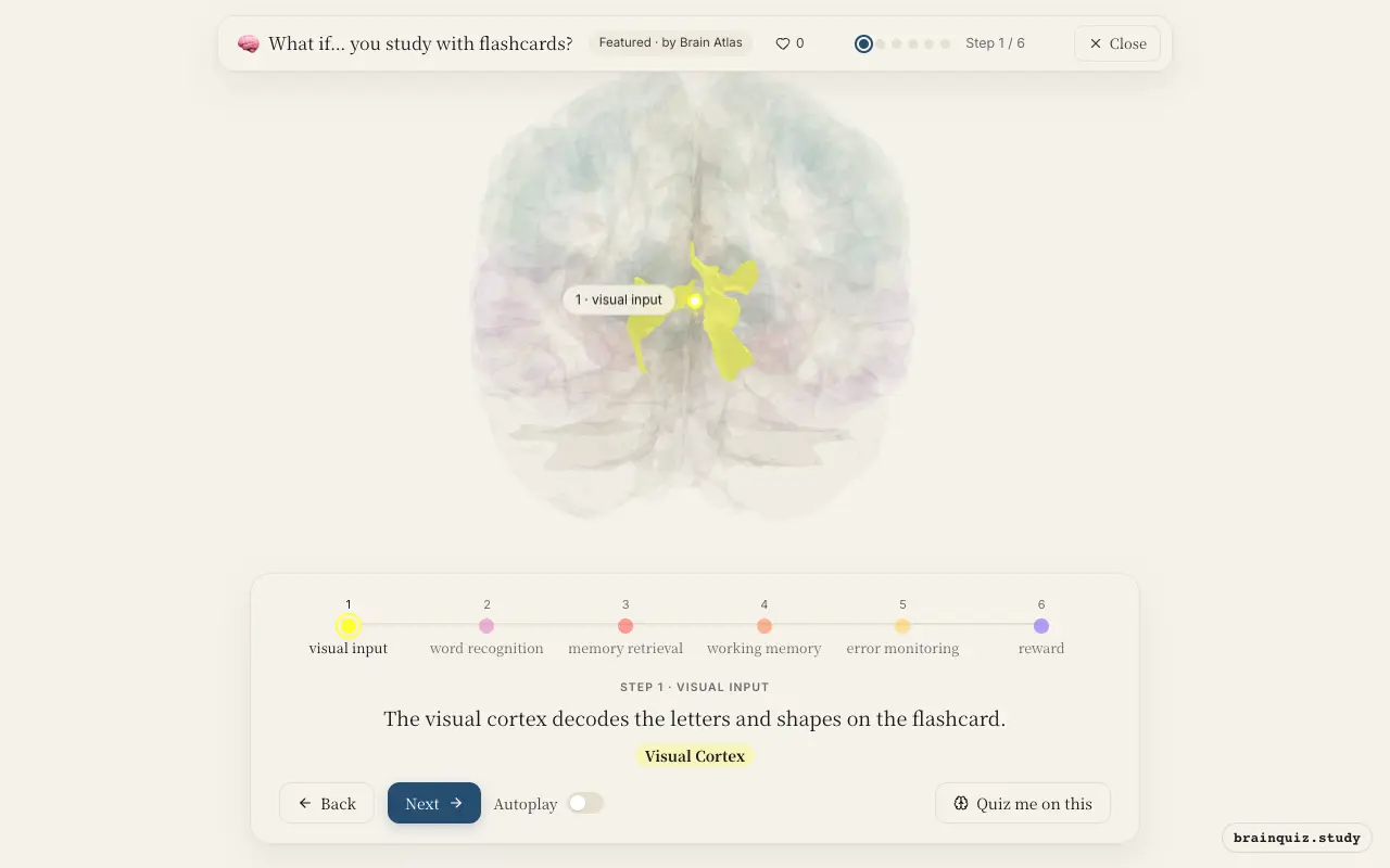
The primary visual cortex in the occipital lobe processes the raw visual information: edges, orientation, and contrast. It passes this to higher visual areas for further analysis. This is the first step in transforming the written word into neural signals.
2. Fusiform Gyrus: word recognition
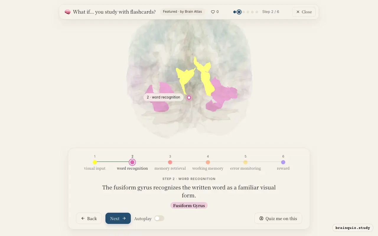
Located in the ventral temporal lobe, the fusiform gyrus contains the visual word form area. It specializes in identifying letter combinations and whole words. This allows the brain to rapidly map the visual input to a known lexical representation.
3. Hippocampus: memory retrieval
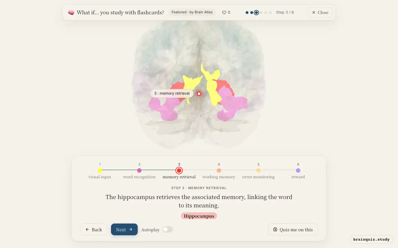
The hippocampus is crucial for retrieving episodic and semantic information. It coordinates the reactivation of cortical patterns that represent the target answer. Repeated retrieval strengthens these connections, improving long-term retention.
4. Prefrontal Cortex: working memory
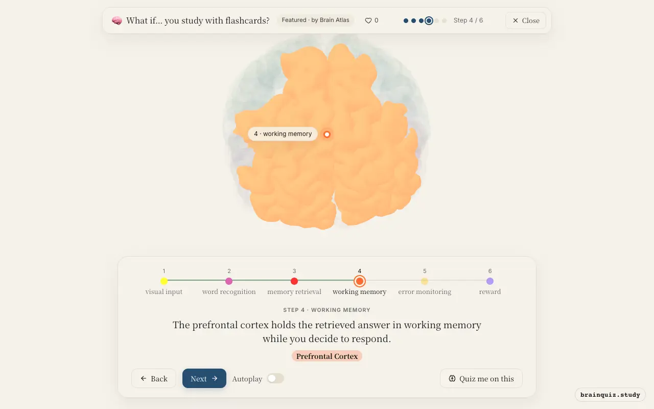
The dorsolateral prefrontal cortex maintains the answer online and supports decision-making. It integrates the retrieved information with the task goal (e.g., 'say the translation'). This region is central to executive control during active recall.
5. Anterior Cingulate Cortex: error monitoring
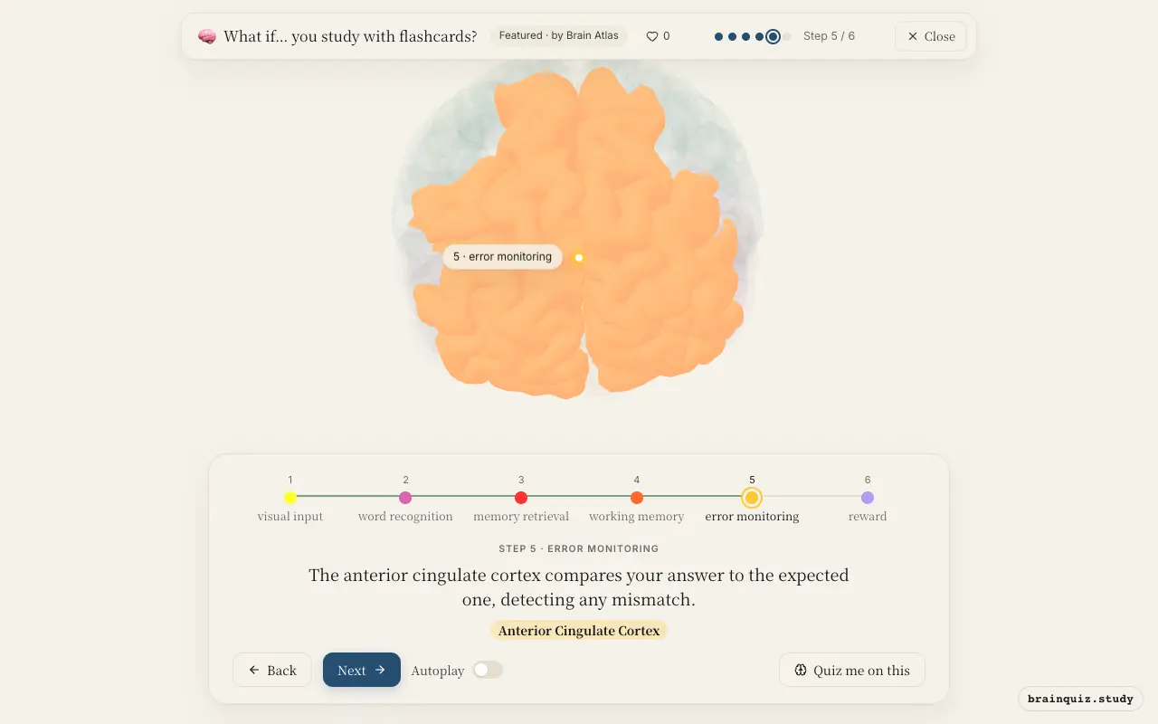
The ACC monitors for conflict between your response and the correct answer. If you make an error, it signals the need for adjustment and heightened attention. Even when correct, it evaluates the outcome, contributing to learning from feedback.
6. Nucleus Accumbens: reward
Correct answers activate the brain's reward circuitry, including the nucleus accumbens. Dopamine release here signals that the response was successful and enhances synaptic plasticity in memory networks. This positive feedback loop motivates continued studying.
Good to know
- Retrieval practice, like using flashcards, strengthens memory more than passive re-reading (the testing effect).
- Spaced repetition schedules optimize long-term retention by timing reviews just before forgetting.
- Dopamine release during correct recall may enhance consolidation of the retrieved memory.
Flashcard recall vs. passive re-reading
During active recall, the hippocampus and prefrontal cortex show greater engagement than during passive reading, which relies more on visual and language areas. Active retrieval produces larger and longer-lasting memory gains, a phenomenon known as the testing effect.
Test yourself
Which brain region is most critical for retrieving the answer when studying with flashcards?
What does the anterior cingulate cortex do during flashcard study?
Which neurotransmitter is released in the nucleus accumbens when you answer correctly?
0 of 3 answered · 0 correct
Simplified: many regions work together at once, and this shows the main players in the order they act.