What happens in your brain when you are grieving?
Updated
Grief triggers the amygdala for emotional salience, anterior cingulate for social pain, insula for visceral feelings, hippocampus for memories, and prefrontal cortex for attempted regulation.
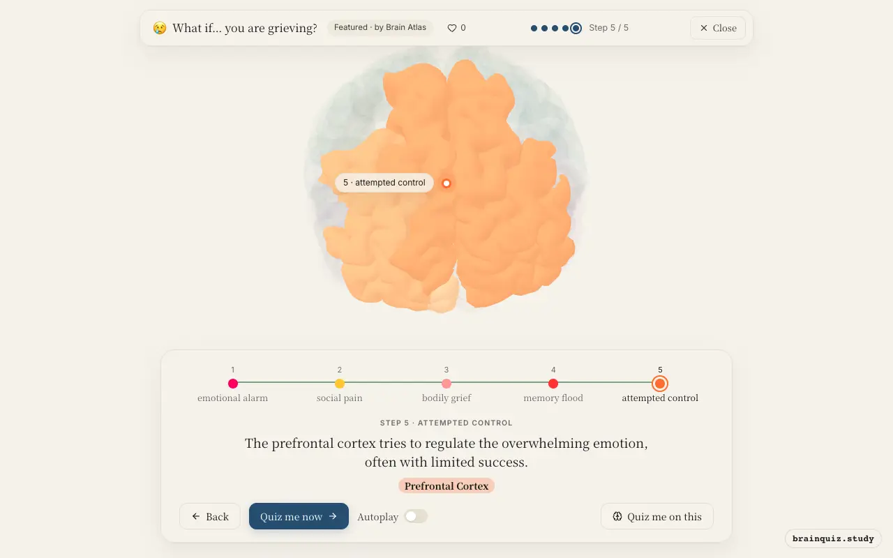
Watch it light up on the 3D brain
Brain regions that light up
| Order | Region | What it does here |
|---|---|---|
| 1 | Amygdala | The amygdala detects the loss as a highly salient emotional event, triggering sadness and distress. |
| 2 | Anterior Cingulate Cortex | The anterior cingulate cortex processes the emotional pain of separation, akin to physical pain. |
| 3 | Insula | The insula processes the visceral feelings of grief, such as heaviness and tears. |
| 4 | Hippocampus | The hippocampus retrieves vivid memories of the deceased, fueling longing and rumination. |
| 5 | Prefrontal Cortex | The prefrontal cortex tries to regulate the overwhelming emotion, often with limited success. |
1. Amygdala: emotional alarm
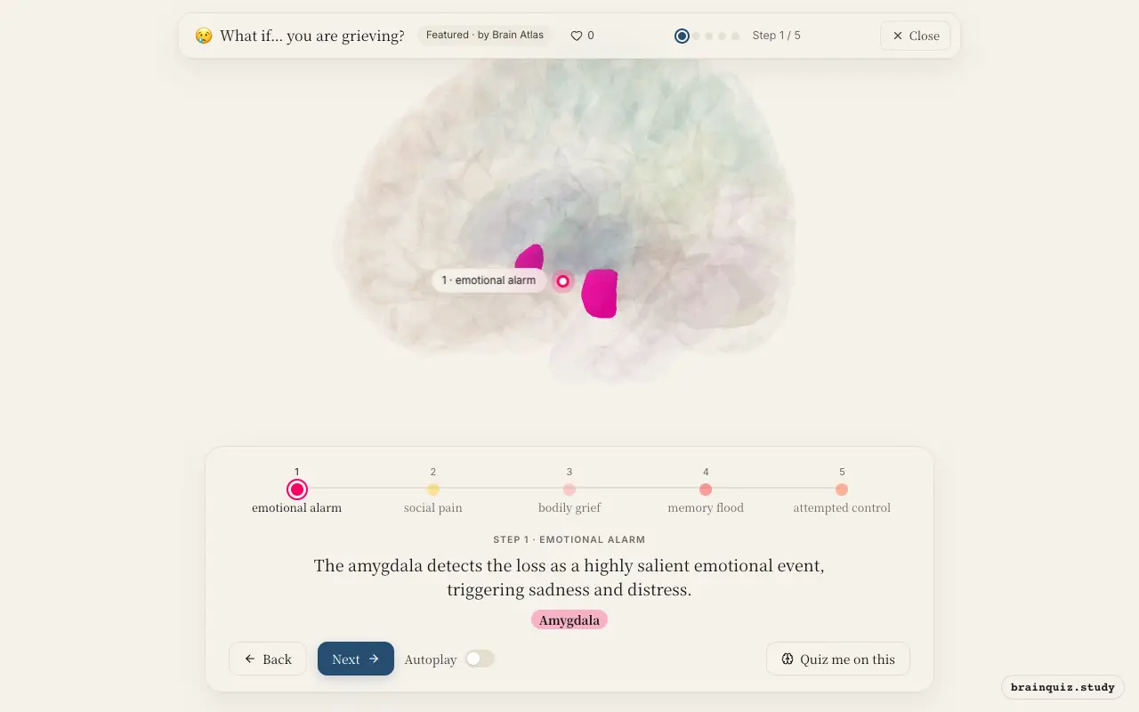
The amygdala rapidly evaluates the emotional significance of the loss, activating the stress response. It biases attention toward grief-related cues and enhances memory consolidation of the event. This initial alarm sets the stage for the intense emotional experience.
2. Anterior Cingulate Cortex: social pain
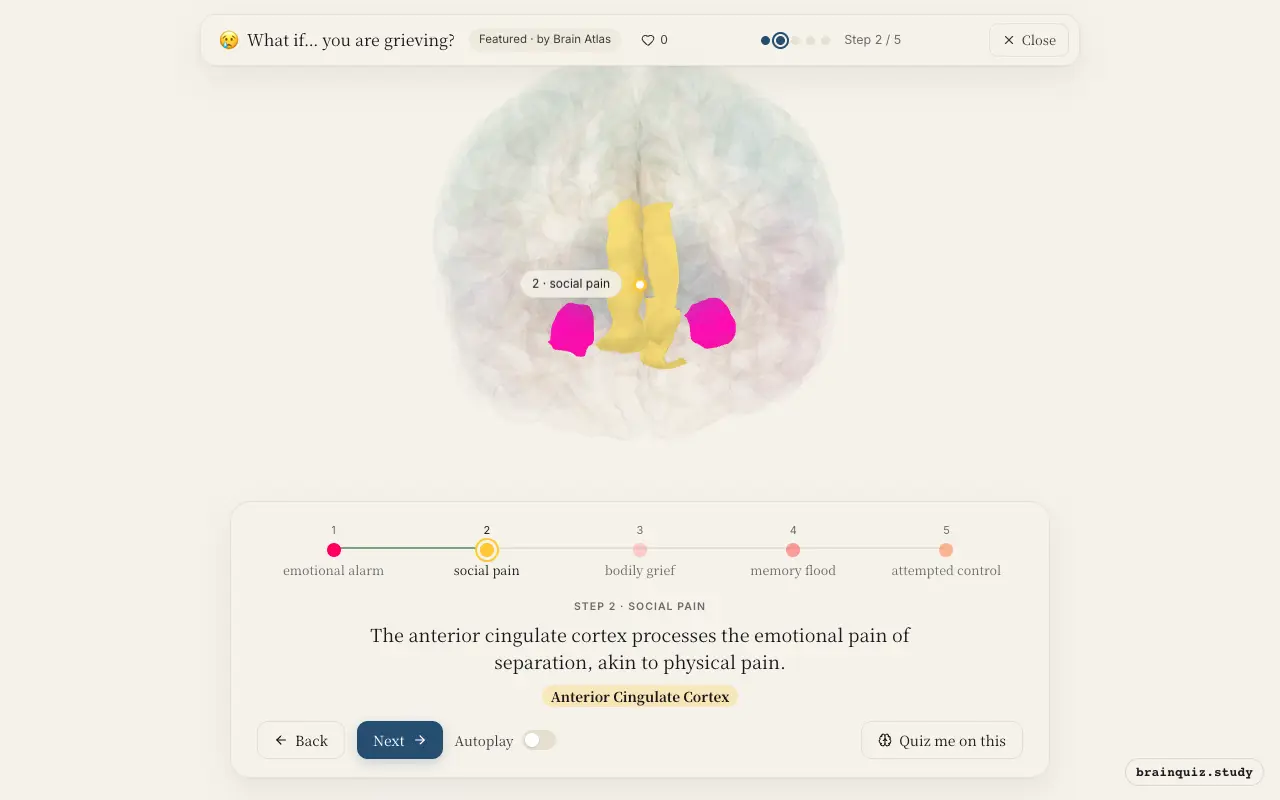
The anterior cingulate cortex is a key node for pain processing, both physical and social. In grief, it responds to the separation from the loved one, registering the distress as painful. This region also monitors conflict between the desire to reunite and reality.
3. Insula: bodily grief
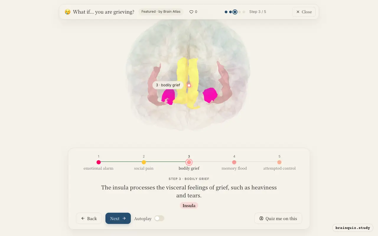
The insula integrates interoceptive signals from the body, representing the physical sensations that accompany grief. It is also involved in empathy and self-awareness, intensifying the feeling of loss. Activation here is thought to contribute to the 'broken heart' sensation.
4. Hippocampus: memory flood
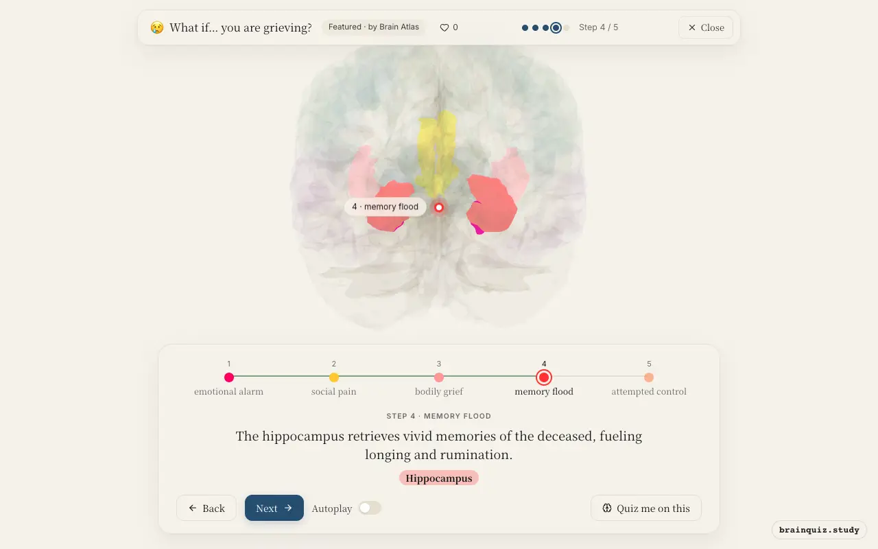
The hippocampus plays a central role in episodic memory, recalling specific moments shared with the lost person. These memories can be triggered by cues and lead to waves of grief. The hippocampus also helps consolidate the reality of the loss into long-term memory.
5. Prefrontal Cortex: attempted control
The prefrontal cortex, especially its dorsolateral and ventromedial parts, is involved in emotion regulation and reappraisal. In grief, it attempts to downregulate the amygdala and insula responses, but chronic stress can impair its function. This leads to difficulty concentrating and decision-making, common in bereavement.
Good to know
- The nucleus accumbens, typically associated with reward, shows altered activity in grief, reflecting the craving for the deceased.
- Grief-related stress can elevate cortisol levels, which over time may impair hippocampal function and memory.
- Some studies suggest that grief and depression share neural features, but grief typically resolves without clinical intervention.
Grief vs. Depression
While grief and depression share overlapping regions like the anterior cingulate and prefrontal cortex, grief is typically triggered by a specific loss and involves intense yearning. Depression often involves more global anhedonia and self-critical thoughts, with less focus on the lost person.
Test yourself
Which brain region is primarily responsible for the intense emotional pain of grief?
The insula's role in grief is best described as:
Which region tries to downregulate the emotional response during grief, but may be overwhelmed?
0 of 3 answered · 0 correct
Simplified: many regions work together at once, and this shows the main players in the order they act.