What happens in your brain when you cry?
Updated
When you cry, the amygdala first flags emotional salience; the anterior cingulate cortex generates the feeling of distress; the hypothalamus triggers autonomic arousal; the brainstem signals lacrimal glands; and the insula maps the bodily changes.
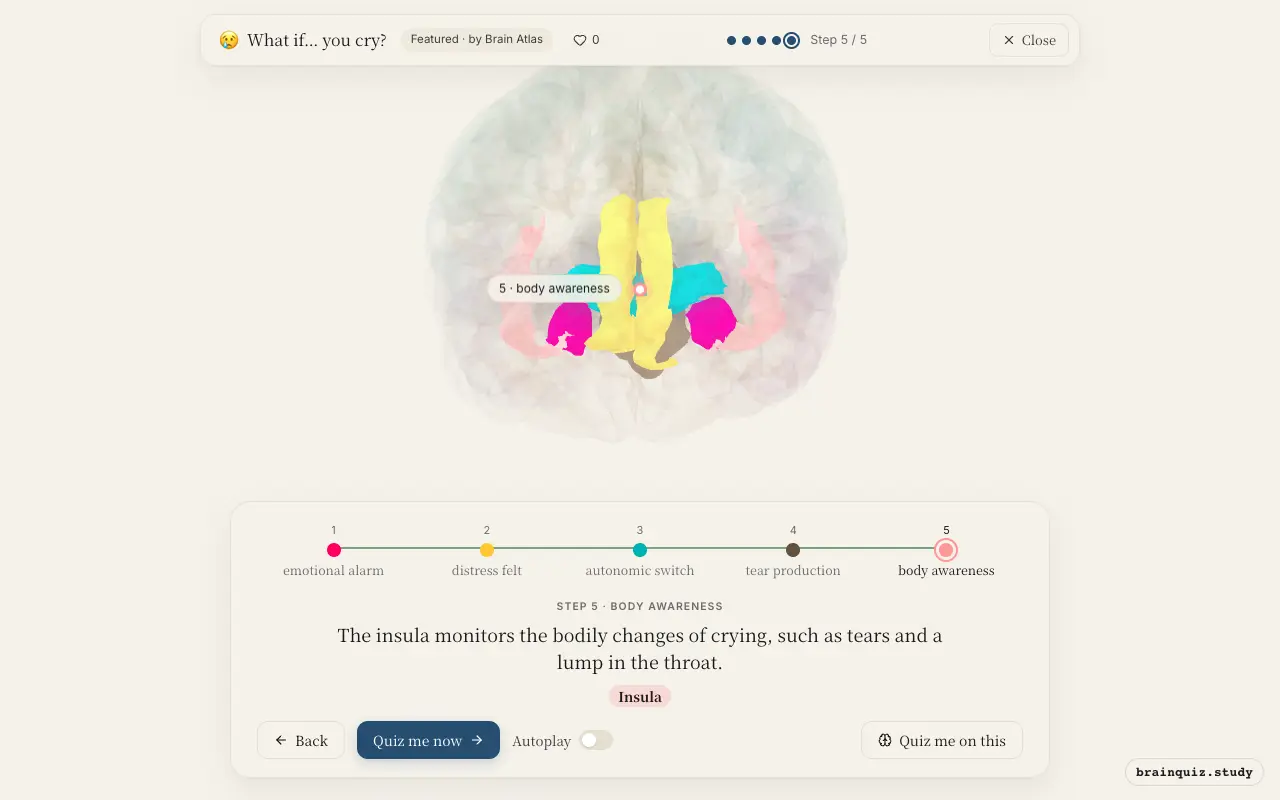
Watch it light up on the 3D brain
Brain regions that light up
| Order | Region | What it does here |
|---|---|---|
| 1 | Amygdala | The amygdala detects the emotional meaning of events and flags them as sad or distressing. |
| 2 | Anterior Cingulate Cortex | The anterior cingulate cortex processes the feeling of distress and the urge to cry. |
| 3 | Hypothalamus | The hypothalamus activates the autonomic nervous system, increasing arousal and preparing the body to cry. |
| 4 | Brainstem | The brainstem sends parasympathetic signals to the lacrimal glands, producing tears, and controls sobbing movements. |
| 5 | Insula | The insula monitors the bodily changes of crying, such as tears and a lump in the throat. |
1. Amygdala: emotional alarm
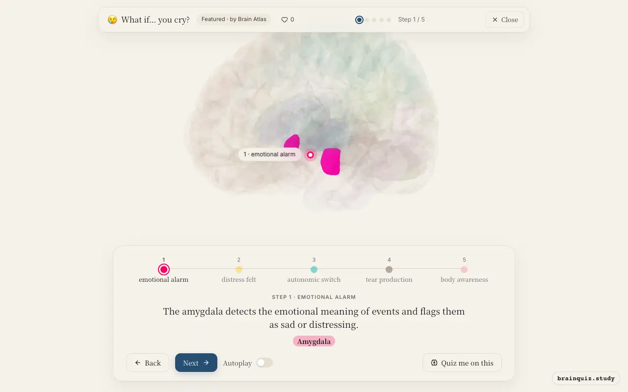
The amygdala is central to processing emotionally salient stimuli. It evaluates sensory input for threat or sadness and triggers rapid, unconscious responses. In crying, its activation is thought to initiate the emotional cascade before conscious awareness.
2. Anterior Cingulate Cortex: distress felt
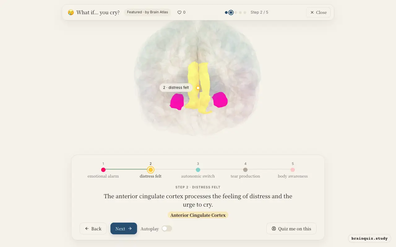
The anterior cingulate cortex (ACC) integrates emotional and bodily information, contributing to the conscious experience of sadness or pain. It is also involved in conflict monitoring, which may reflect the internal struggle when trying not to cry.
3. Hypothalamus: autonomic switch
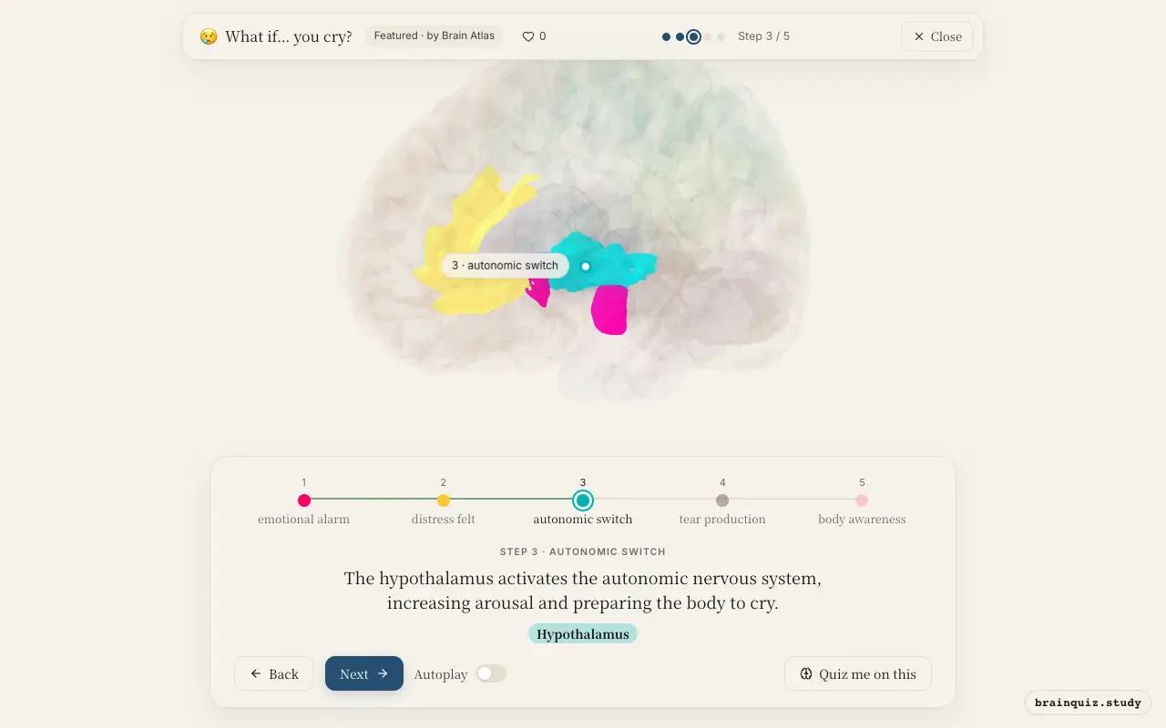
The hypothalamus is a key control center for autonomic and endocrine responses. During emotional crying, it coordinates sympathetic activation (e.g., increased heart rate) and parasympathetic outflow to the lacrimal glands. It may also trigger hormone release, such as adrenocorticotropic hormone.
4. Brainstem: tear production
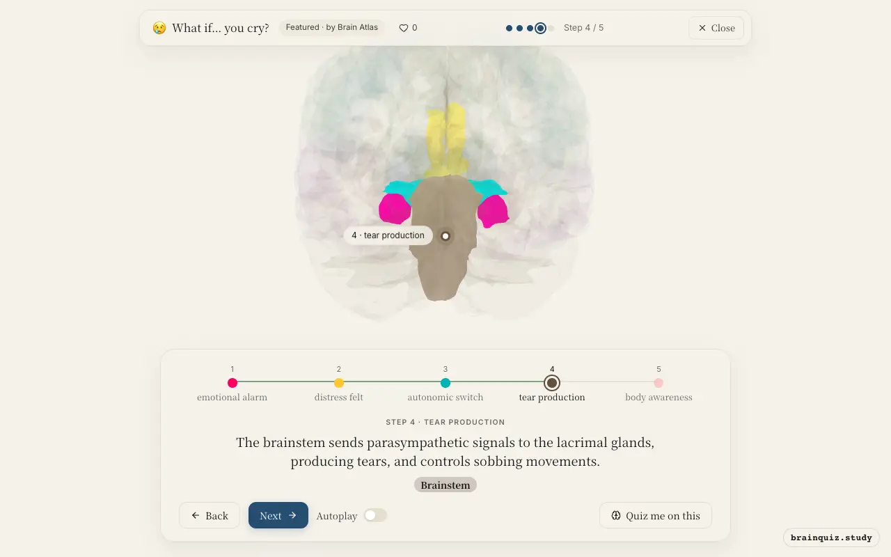
Nuclei in the brainstem, including the superior salivatory nucleus, provide parasympathetic innervation to the lacrimal glands via the facial nerve. Brainstem respiratory centers also generate the irregular breathing pattern of sobbing. This is the final common pathway for tear secretion.
5. Insula: body awareness
The insula integrates interoceptive signals from the body, giving rise to the felt experience of the physical changes during crying. It contributes to self-awareness and empathy, linking the internal state to the emotional feeling.
Good to know
- Studies suggest that emotional tears contain higher levels of stress hormones and endorphins than reflex tears, possibly aiding in stress relief.
- Crying often begins with sympathetic arousal (increased heart rate) and is followed by parasympathetic activation, which may help restore calm.
- Crying has a strong social component; people often cry in the presence of others, and tears can signal distress and elicit support.
emotional crying vs reflex tears
Emotional crying involves the amygdala, anterior cingulate, and hypothalamus, reflecting a limbic response. Reflex tears, like from onion fumes, are triggered by irritation of the cornea, processed mainly by the trigeminal nerve and brainstem, with little to no limbic involvement.
Test yourself
Which brain region is primarily responsible for detecting the emotional significance of a sad event, triggering the crying response?
What is the main role of the hypothalamus in crying?
Which sequence best describes the neural pathway of emotional crying?
0 of 3 answered · 0 correct
Simplified: many regions work together at once, and this shows the main players in the order they act.