What happens in your brain when you get a concussion?
Updated
A concussion causes rotational shearing of axons. The brainstem's arousal system can fail, sometimes causing loss of consciousness; the thalamus scrambles sensory relay; and the prefrontal cortex and hippocampus impair thinking and memory.
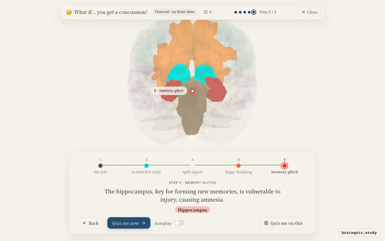
Watch it light up on the 3D brain
Brain regions that light up
| Order | Region | What it does here |
|---|---|---|
| 1 | Brainstem | Rotational forces stretch the brainstem, disrupting the reticular activating system that keeps you awake. |
| 2 | Thalamus | The thalamus, the brain's sensory relay, becomes overloaded and fails to properly route information. |
| 3 | Corpus Callosum | Shearing forces stretch the corpus callosum, the bridge between hemispheres, slowing communication. |
| 4 | Prefrontal Cortex | The prefrontal cortex, responsible for executive function, shows reduced activity, causing brain fog. |
| 5 | Hippocampus | The hippocampus, key for forming new memories, is vulnerable to injury, causing amnesia. |
1. Brainstem: the jolt
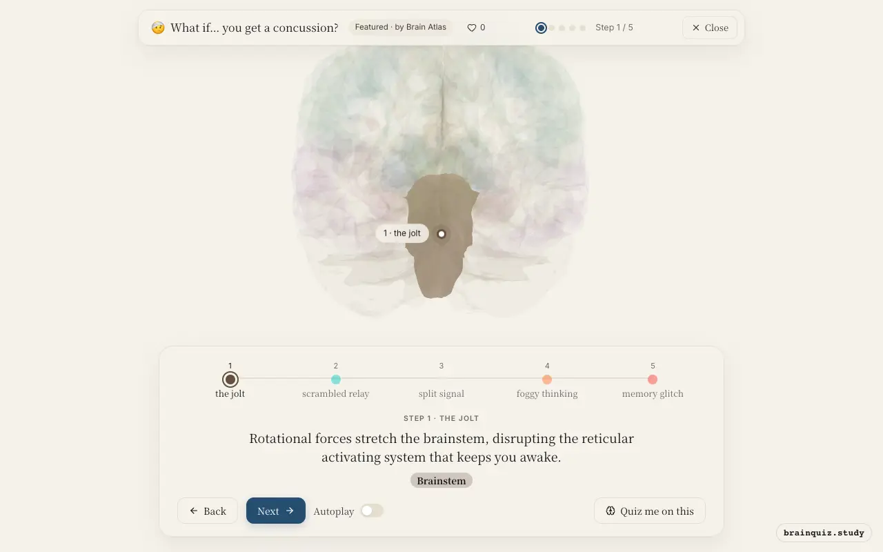
The brainstem houses the reticular formation, which maintains wakefulness. Concussive forces can briefly disrupt this network, leading to loss of consciousness or a feeling of being dazed. Feeling dazed is common, but loss of consciousness is rare.
2. Thalamus: scrambled relay
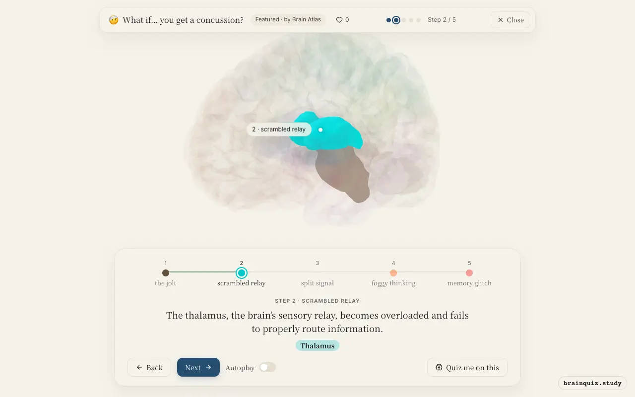
The thalamus integrates and relays sensory and motor signals to the cortex. After a concussion, its widespread connections are disrupted, contributing to confusion, slowed reaction times, and difficulty focusing attention. This is part of the metabolic crisis that follows injury.
3. Corpus Callosum: split signal
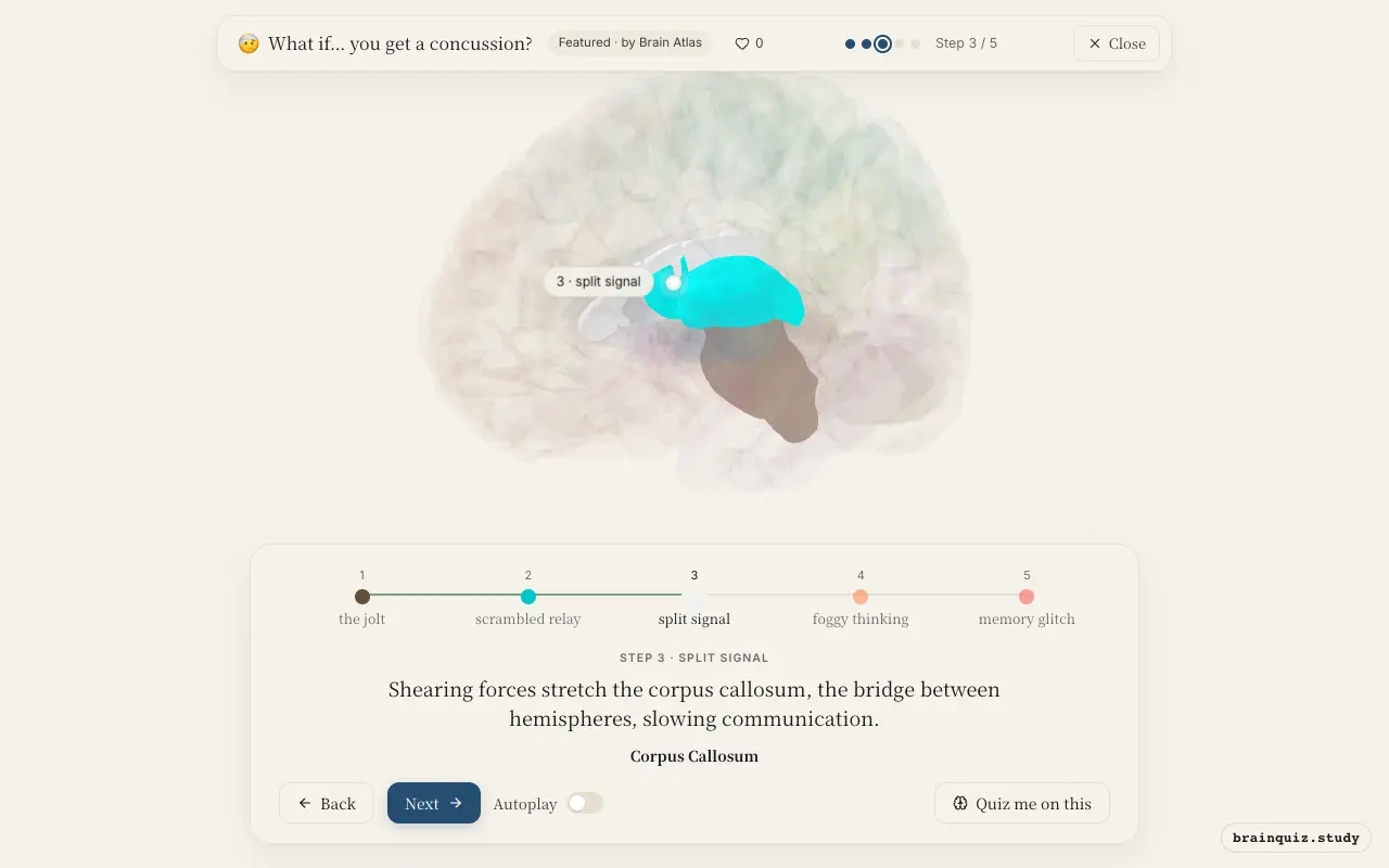
The corpus callosum consists of millions of axons connecting the left and right hemispheres. It is especially vulnerable to rotational injury because it sits in the midline. Damage here can cause subtle cognitive deficits, like slower processing speed and difficulty with tasks requiring both hemispheres.
4. Prefrontal Cortex: foggy thinking
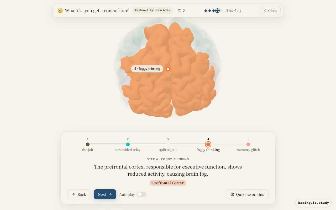
The prefrontal cortex manages working memory, attention, and decision-making. Concussion impairs its function due to axonal injury and energy deficits, leading to difficulty concentrating, mental fatigue, and slowed problem-solving. These symptoms often persist during recovery.
5. Hippocampus: memory glitch
The hippocampus is essential for encoding new episodic memories. Concussion can disrupt its function through excitotoxicity and metabolic stress, leading to anterograde amnesia—difficulty remembering events after the injury. Usually this resolves, but repeated injuries may cause lasting damage.
Good to know
- Concussion involves a metabolic cascade of ionic flux and glutamate release, not macroscopic bleeding.
- Standard CT or MRI scans usually appear normal after concussion because the injury is at the cellular level.
- Loss of consciousness occurs in fewer than 10% of concussions, so most people remain awake.
Concussion vs. Whiplash
While both can occur after a sudden head movement, concussion involves brain dysfunction from axonal shearing, seen as disrupted activity in the brainstem and cortex. Whiplash primarily injures neck muscles and ligaments, with brain activity largely normal. Symptoms overlap—headache, dizziness—but whiplash lacks the cognitive and arousal deficits of concussion.
Test yourself
Which brain region's disruption most directly causes loss of consciousness in a concussion?
Why do standard CT scans often appear normal after a concussion?
Which symptom is most directly linked to hippocampal dysfunction after concussion?
0 of 3 answered · 0 correct
Simplified: many regions work together at once, and this shows the main players in the order they act. For learning only, not medical advice.