What happens in your brain when you get Parkinson's disease?
Updated
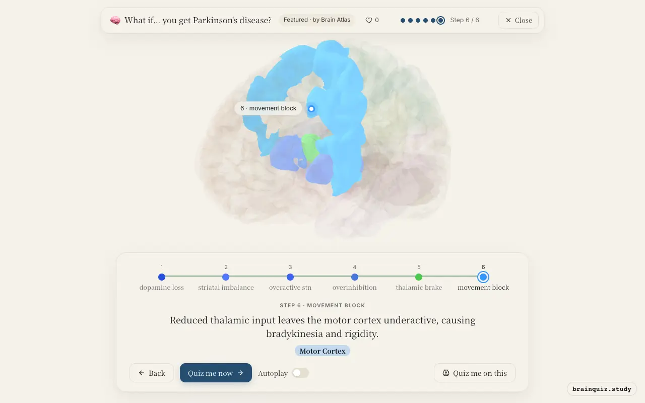
In Parkinson's, dopamine neurons in the substantia nigra die, reducing dopamine to the putamen. This overactivates the subthalamic nucleus and internal globus pallidus, which overly inhibits the thalamus, weakening motor cortex drive to muscles, causing slowness and rigidity.
Watch it light up on the 3D brain
Brain regions that light up
| Order | Region | What it does here |
|---|---|---|
| 1 | Substantia Nigra | Dopamine-producing neurons in the substantia nigra degenerate. |
| 2 | Putamen | Reduced dopamine disrupts the putamen's control of basal ganglia pathways. |
| 3 | Subthalamic Nucleus | The subthalamic nucleus becomes hyperactive due to loss of inhibition. |
| 4 | Globus Pallidus, internal segment | The internal globus pallidus fires excessively, inhibiting the thalamus. |
| 5 | Ventral Anterior Nucleus | The thalamus is overly inhibited, reducing its excitatory drive to the motor cortex. |
| 6 | Motor Cortex | Reduced thalamic input leaves the motor cortex underactive, causing bradykinesia and rigidity. |
1. Substantia Nigra: dopamine loss
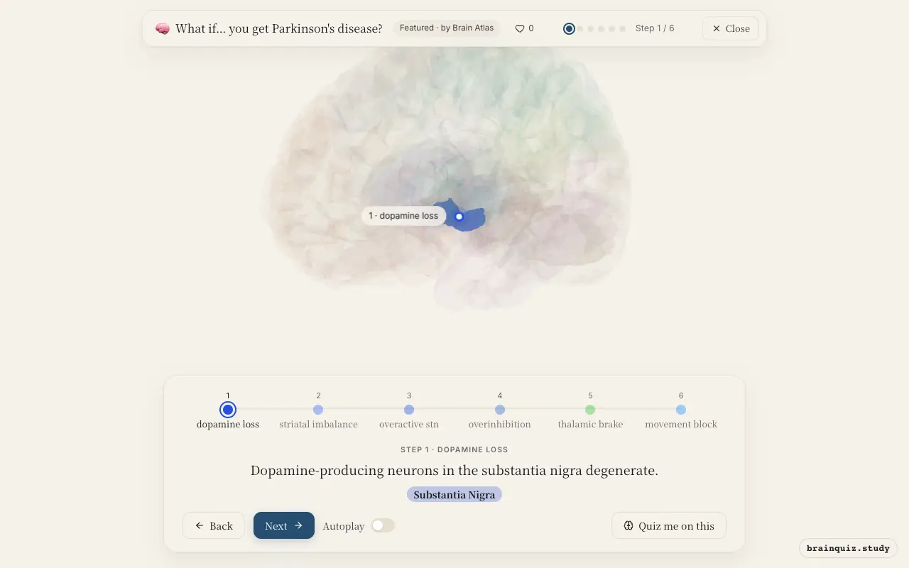
The substantia nigra pars compacta normally sends dopamine to the striatum. In Parkinson's, these neurons die, causing profound dopamine deficiency. This loss is the pathological hallmark of the disease.
2. Putamen: striatal imbalance
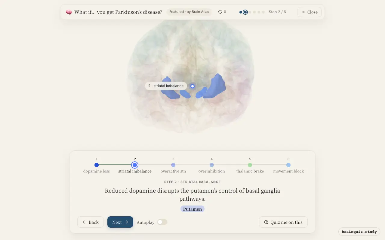
The putamen is the main input nucleus of the basal ganglia receiving nigral dopamine. Without dopamine, the direct pathway is underactive and the indirect pathway overactive, leading to abnormal circuit output.
3. Subthalamic Nucleus: overactive stn
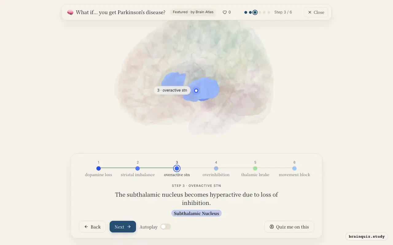
Normally, the external globus pallidus inhibits the subthalamic nucleus, but in Parkinson's that inhibition is reduced. This leads to excessive excitatory output from the STN, driving the internal globus pallidus.
4. Globus Pallidus, internal segment: overinhibition
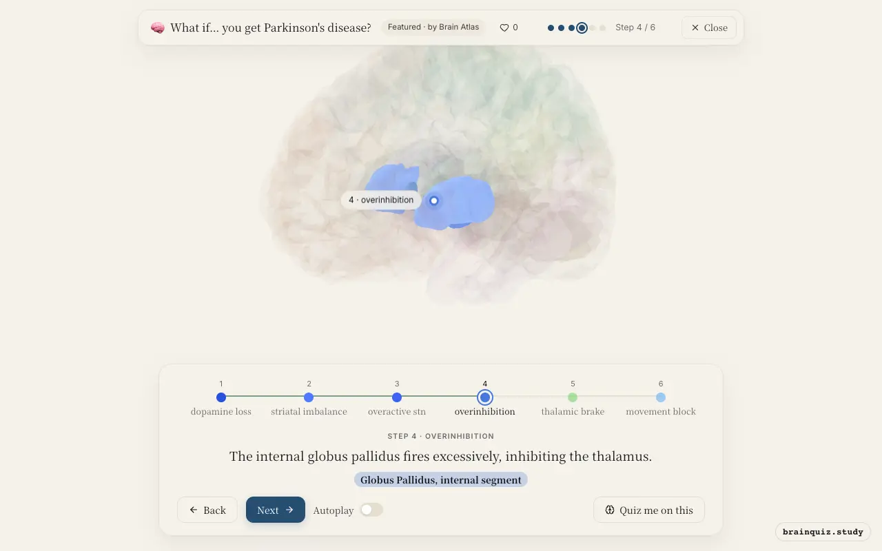
The GPi is the main basal ganglia output nucleus. In Parkinson's, its neurons become overactive and fire in abnormal patterns, sending strong inhibitory signals to the thalamus.
5. Ventral Anterior Nucleus: thalamic brake
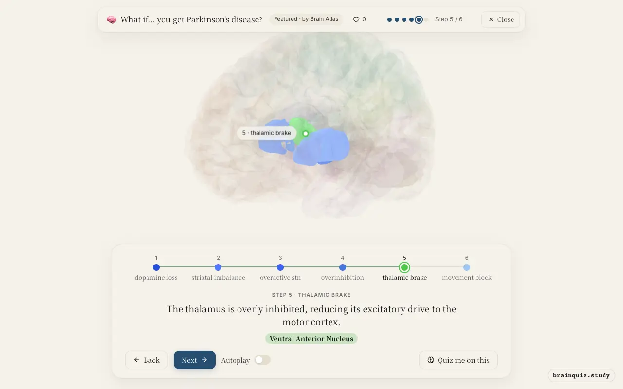
The ventral anterior thalamic nucleus relays basal ganglia output to the frontal cortex. When GPi inhibition is too strong, the thalamus cannot properly excite the motor cortex, impairing movement initiation.
6. Motor Cortex: movement block
The motor cortex relies on thalamic drive to plan and execute voluntary movements. With less excitation, commands to muscles are delayed and weak, producing the slow, stiff movements typical of Parkinson's.
Good to know
- The substantia nigra pars compacta normally supplies dopamine to the putamen via the nigrostriatal pathway.
- Levodopa, a dopamine precursor, is a common treatment because it can cross the blood-brain barrier.
- Deep brain stimulation of the subthalamic nucleus can improve motor symptoms in advanced Parkinson's.
Parkinson's vs. Huntington's disease
In Parkinson's, dopamine loss overactivates the indirect pathway, causing too much inhibition of the thalamus and reduced movement. In Huntington's, degeneration of striatal neurons in the indirect pathway reduces inhibition of the GPe, ultimately reducing excitation of the GPi, leading to excessive thalamic excitation and chorea. Thus, opposite motor symptoms arise from opposite basal ganglia dysfunctions.
Test yourself
Which brain region shows the primary neuronal loss in Parkinson's?
How does Parkinson's affect the internal globus pallidus?
Which treatment targets the subthalamic nucleus to reduce symptoms?
0 of 3 answered · 0 correct
Simplified: many regions work together at once, and this shows the main players in the order they act. For learning only, not medical advice.