Motor Cortex — Function, Location & Quiz
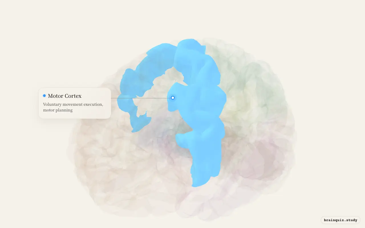
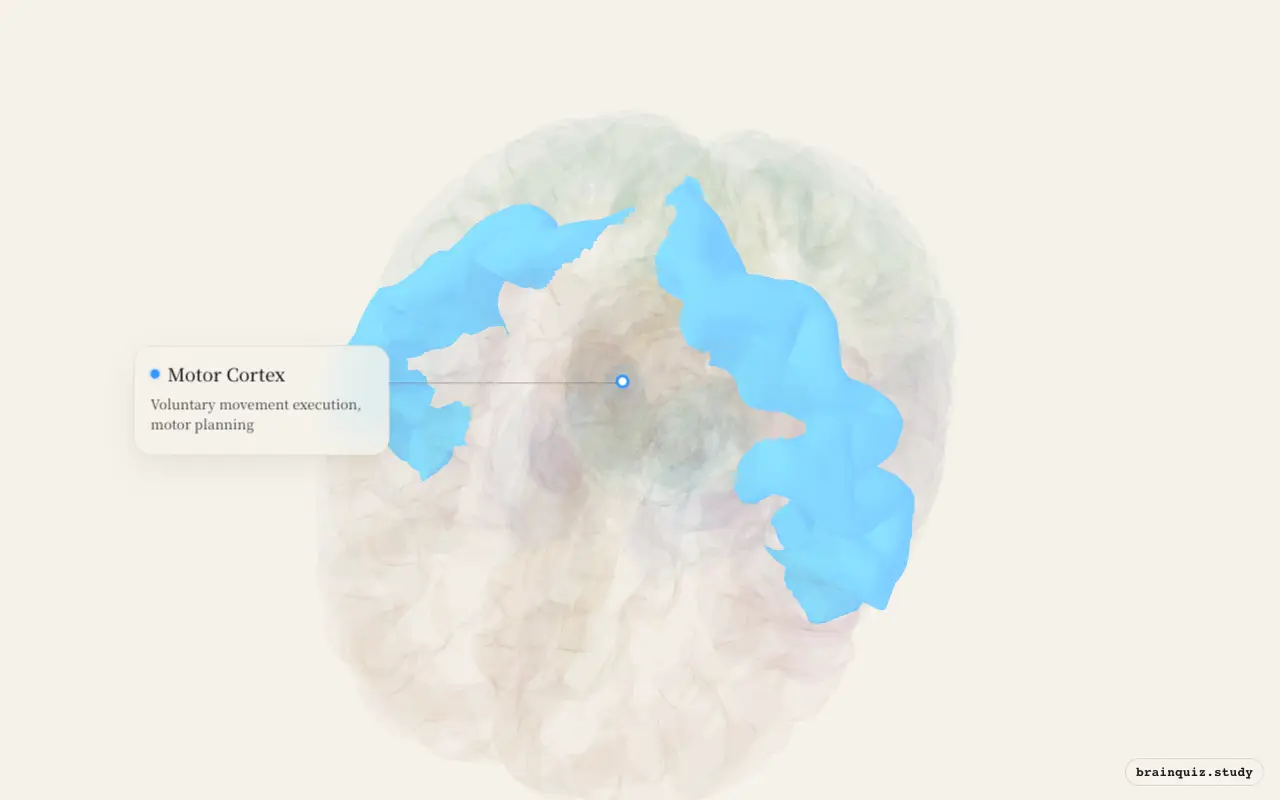
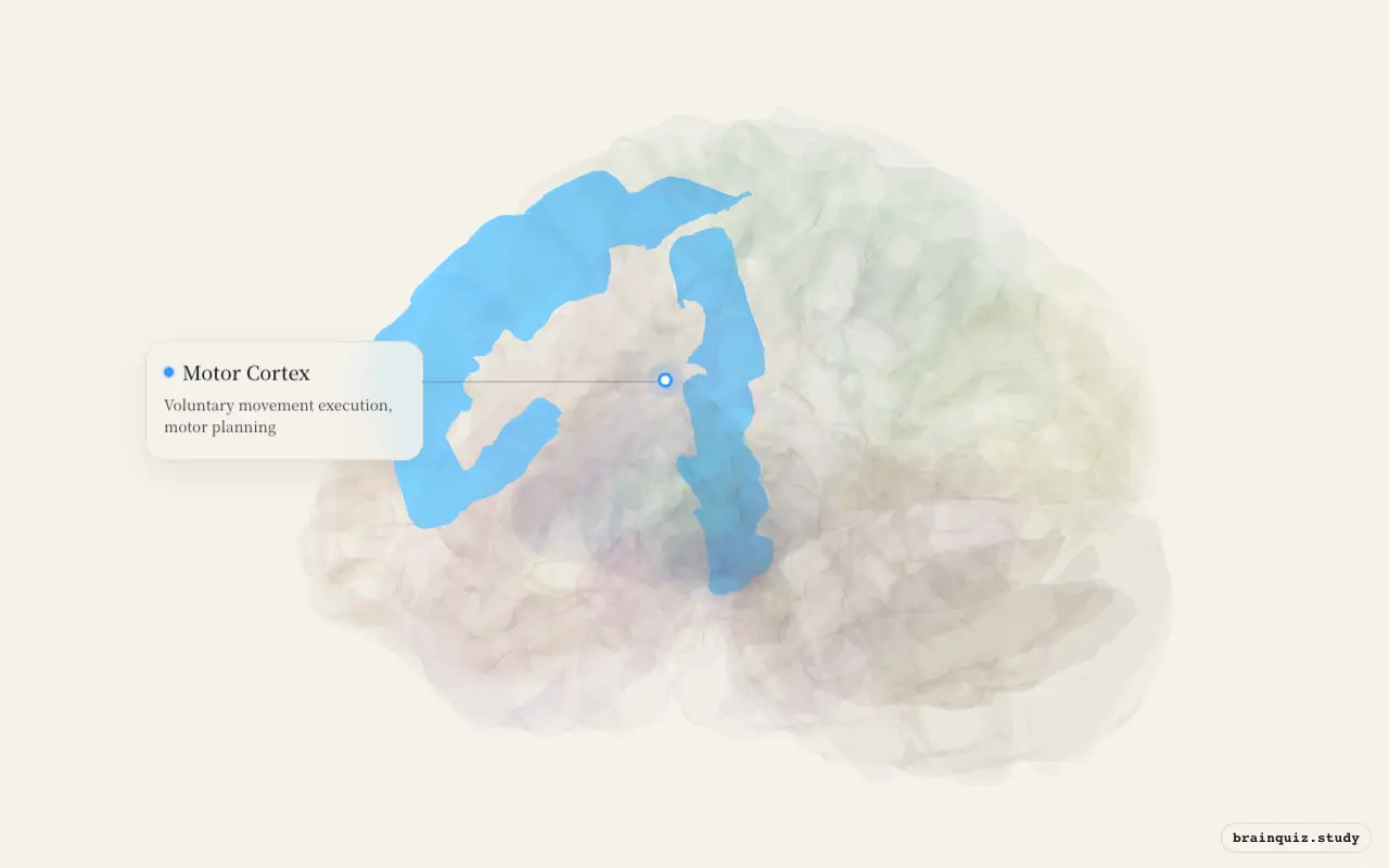
Primary motor cortex is the narrow strip where a movement plan becomes descending command. Its position on the precentral gyrus is the landmark to fix first, because the almost mirror-like postcentral gyrus just behind it is sensory rather than motor.
The motor homunculus turns this strip into a clinical map: a small cortical lesion can weaken a particular contralateral body region, and vascular territory predicts which part is most exposed. That link from folded surface to bedside weakness is why the region appears so often in localization questions.
Test this anatomy in the Lesion Localization Quiz: 15 Clinical Questions or explore it on the interactive 3D brain.
Where is the Motor Cortex located?
Voluntary movement execution, motor planning.
What does the Motor Cortex do?
- Direct control of voluntary movements via corticospinal tract
- Somatotopic organization — motor homunculus
- Force and direction coding of movements
- Motor learning and adaptation
Motor Cortex pathways and connections
- Corticospinal tract → spinal motor neurons (direct pathway)
- M1 ← Premotor cortex, SMA (movement planning)
- M1 ← Cerebellum via thalamus (motor correction)
- M1 ← Basal ganglia via thalamus (movement initiation)
What happens when the Motor Cortex is damaged?
- Lesion → contralateral hemiparesis or hemiplegia
- Upper motor neuron signs: spasticity, hyperreflexia, Babinski sign
- Stroke in MCA territory often affects face and arm (lateral homunculus)
- Penfield's stimulation studies mapped the motor homunculus
Motor Cortex key facts
Atlas category: cortical. Brodmann areas or landmark: BA 4 (primary motor cortex, M1).
- Motor homunculus: hands and face have disproportionately large representation
- Contains giant Betz cells — among the largest neurons in the CNS
- Penfield & Boldrey (1937) — mapped the homunculus during epilepsy surgery
- M1 uses population coding — movement direction encoded by population vector
Motor Cortex exam tip
Motor homunculus = upside down, with feet at the medial surface (ACA territory) and face lateral (MCA territory). Know the vascular implications!
Common questions
Where is the primary motor cortex?
It lies in the precentral gyrus of the frontal lobe, immediately anterior to the central sulcus. It corresponds mainly to Brodmann area 4.
What does the motor cortex control?
It sends commands for voluntary movement through descending pathways, especially the corticospinal tract. Its body map gives the hands and face disproportionately large cortical representation.
What are the signs of a motor cortex lesion?
A lesion can cause contralateral weakness with upper-motor-neuron signs such as spasticity, hyperreflexia and a Babinski response. The affected body part depends on the lesion’s place on the motor homunculus.
When does the Motor Cortex light up?
- 😂 When you laugh at a joke: The motor cortex initiates the coordinated muscle contractions of laughter.
- 💩 When you poop: The motor cortex sends signals to relax the external anal sphincter and increase abdominal pressure.
- 💧 When you get thirsty: The motor cortex initiates the movements to obtain and drink water.
- 🥱 When you yawn: Brainstem motor circuits command jaw, face, and respiratory muscles to produce the yawn.
- 🫁 When you hold your breath: The motor cortex sends signals to inhibit the diaphragm and chest muscles.
- 😂 When you get tickled: The motor cortex initiates voluntary squirming and withdrawal movements.
- ✋ When you get an itch: The motor cortex initiates the scratching movement.
- 🦵 When your leg falls asleep: The motor cortex plans and executes movements to relieve the pressure.