What happens in your brain when your leg falls asleep?
Updated
When your leg falls asleep, the thalamus relays erratic nerve signals to the somatosensory cortex, which interprets them as tingling. The insula makes you aware of the odd feeling, and the anterior cingulate flags discomfort, prompting the motor cortex to move your leg.
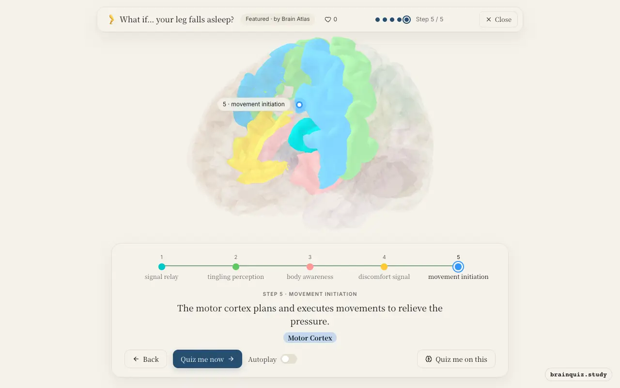
Watch it light up on the 3D brain
Brain regions that light up
| Order | Region | What it does here |
|---|---|---|
| 1 | Thalamus | The thalamus relays distorted sensory signals from the leg to the cortex. |
| 2 | Somatosensory Cortex | The somatosensory cortex interprets the signals as tingling or numbness. |
| 3 | Insula | The insula generates a conscious awareness of the odd bodily state. |
| 4 | Anterior Cingulate Cortex | The anterior cingulate flags the sensation as unpleasant and in need of action. |
| 5 | Motor Cortex | The motor cortex plans and executes movements to relieve the pressure. |
1. Thalamus: signal relay
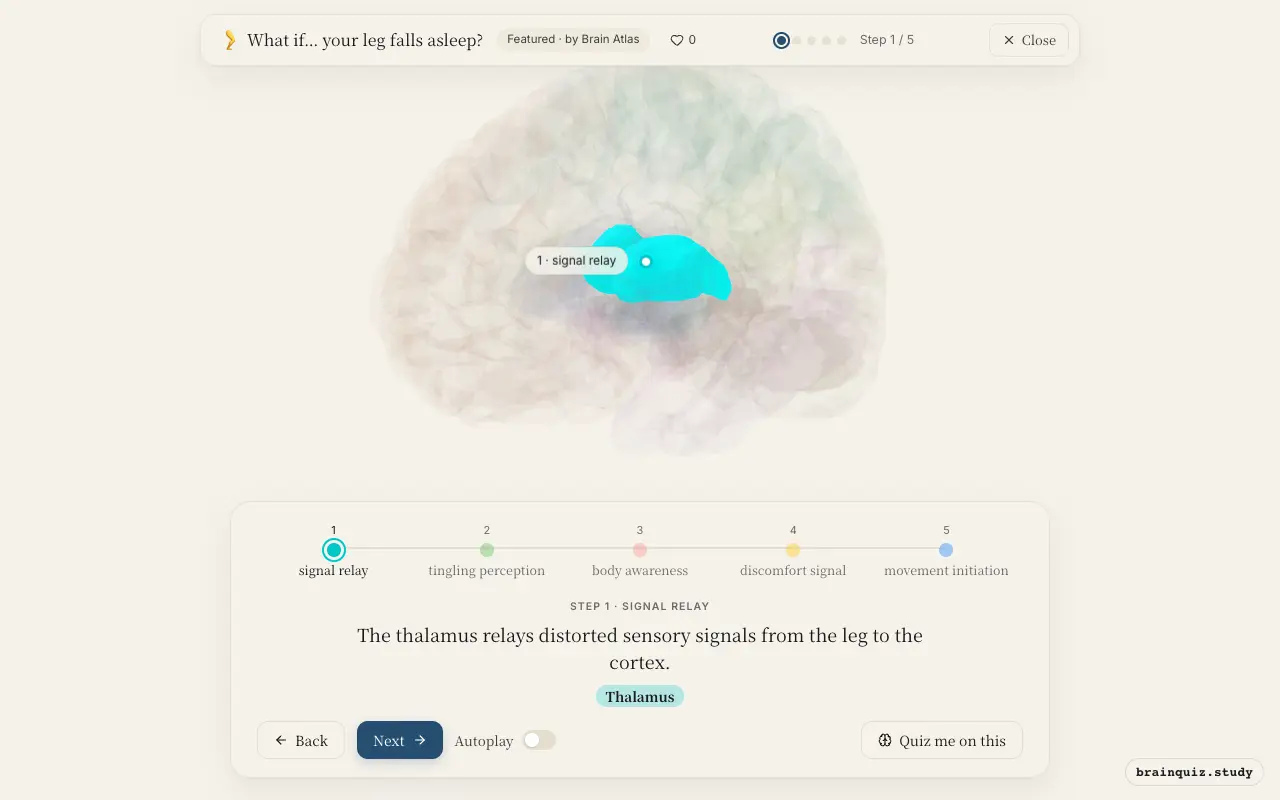
The thalamus acts as a gateway for sensory information, forwarding nerve impulses from the compressed leg nerve. The signals may be irregular because the nerve is firing abnormally. This relay is the first step in the brain's processing of the sensation.
2. Somatosensory Cortex: tingling perception
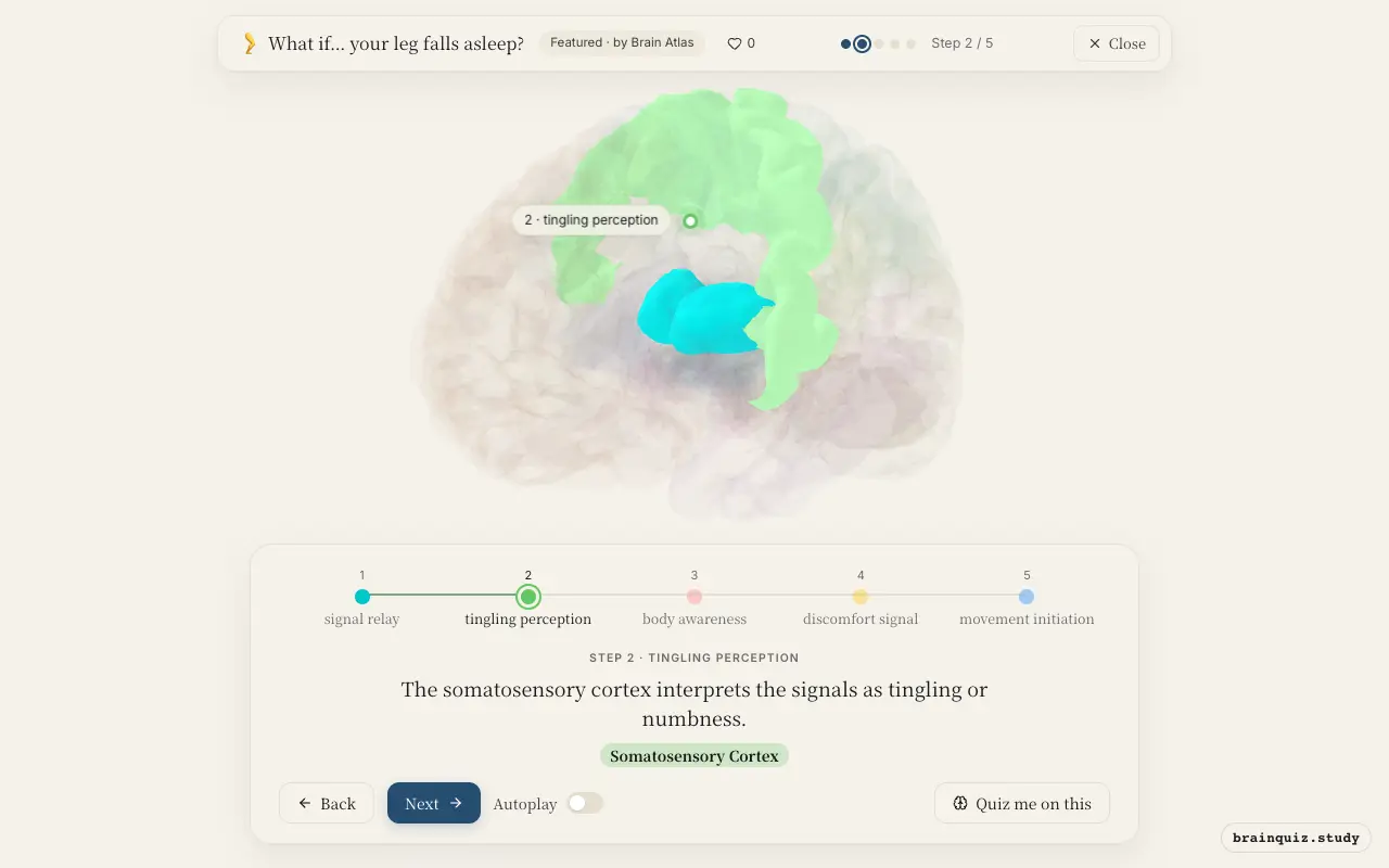
Located in the parietal lobe, the somatosensory cortex maps touch and proprioception from the body. Since the incoming signals are unusual, it creates the perception of pins and needles rather than normal touch. This misinterpretation is what you consciously feel.
3. Insula: body awareness
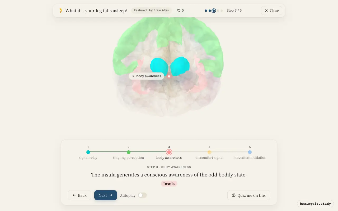
The insula integrates internal body signals to create interoception, the sense of what is happening inside your body. It signals that the leg feels 'different' or 'asleep', contributing to the strange subjective experience. This awareness motivates you to pay attention to the leg.
4. Anterior Cingulate Cortex: discomfort signal
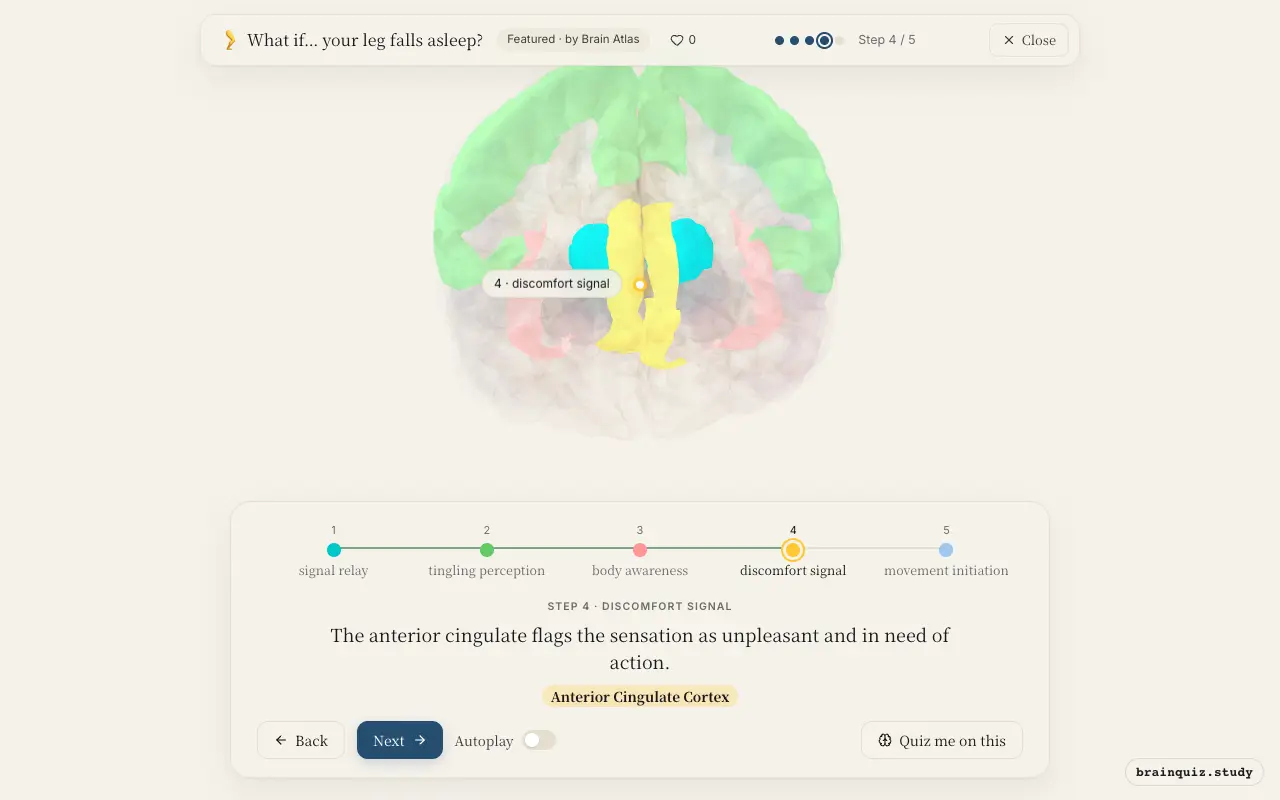
The anterior cingulate cortex is involved in error detection and pain processing. It evaluates the mismatch between the expected normal leg sensation and the actual tingling, producing a sense of discomfort or urgency. This drives the motivation to change position.
5. Motor Cortex: movement initiation
Once you decide to move, the motor cortex sends commands to leg muscles, such as shaking or repositioning. This action alleviates the nerve compression and restores normal blood flow, ending the paresthesia.
Good to know
- The tingling sensation (paresthesia) arises from temporary compression of a nerve, often the sciatic or peroneal nerve, not from brain activity itself.
- Even though the leg feels 'asleep', the brain's somatosensory cortex remains active, but it receives a barrage of spontaneous signals due to nerve ischemia.
- Shifting position relieves pressure on the nerve, and normal sensation returns within minutes as blood flow is restored.
Paresthesia vs normal touch
When you actually touch your sleeping leg, the touch signal may be reduced or distorted, so the somatosensory cortex responds less to external pressure. Instead, spontaneous firing from the compressed nerve produces tingling without any external stimulus, so the brain's activity reflects internal noise rather than accurate touch.
Test yourself
Which brain region first receives sensory signals from the leg before they reach the cortex?
What is the primary role of the somatosensory cortex in this scenario?
Why might the anterior cingulate cortex become active?
0 of 3 answered · 0 correct
Simplified: many regions work together at once, and this shows the main players in the order they act.