What Is the Sensorimotor Network? Brain Regions and Function
Updated
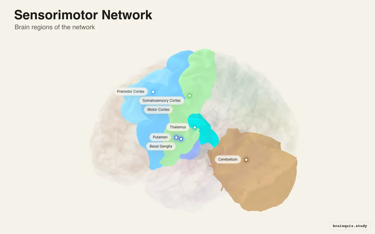
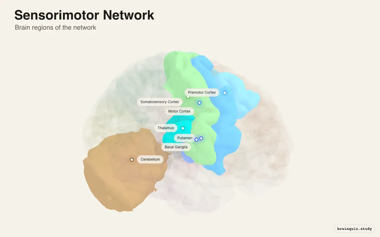
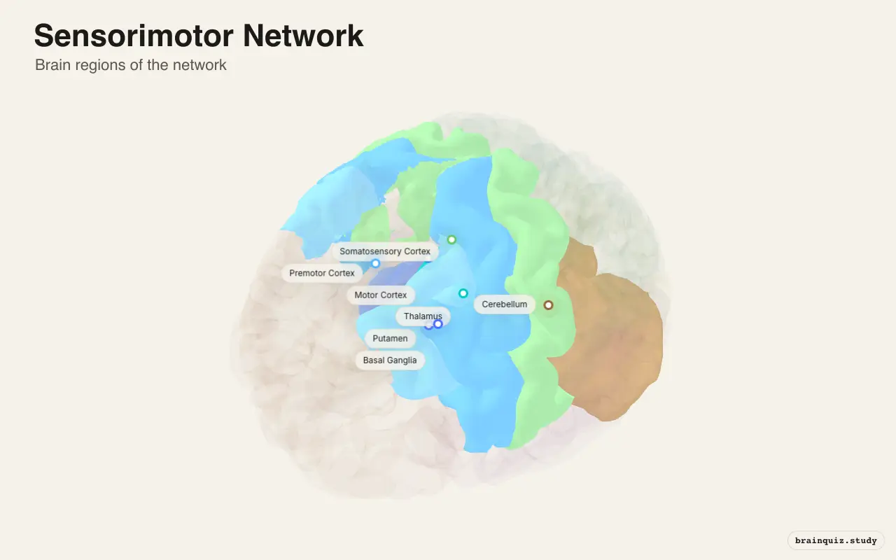
The Sensorimotor Network is a set of brain regions, including the motor cortex, somatosensory cortex, premotor cortex, and subcortical loops, that coordinates voluntary movement and integrates sensory feedback.
The sensorimotor network was identified through resting-state functional connectivity MRI in the early 2000s, revealing coherent spontaneous activity between motor and sensory areas. It is fundamental for understanding how the brain plans, executes, and adapts movements, and its dysfunction underlies many motor disorders.
Which brain regions are in the Sensorimotor Network?
- Motor Cortex: Executes voluntary movements by sending commands to the spinal cord and brainstem; encodes force and direction of movement.
- Somatosensory Cortex: Processes touch, proprioception, and pain; provides real-time feedback about body position and contact.
- Premotor Cortex: Plans movement sequences and integrates sensory cues; includes the supplementary motor area for bimanual coordination.
- Putamen: Part of the basal ganglia; regulates movement initiation and scaling through dopamine-dependent striatal circuits.
- Cerebellum: Compares intended movement with sensory feedback; fine-tunes timing and coordination, enabling motor learning.
- Thalamus: Relays motor and sensory signals between cortex and subcortical structures; gates information flow in motor loops.
- Basal Ganglia: Subcortical nuclei that select and inhibit competing motor programs; essential for habit formation and action selection.
What does the Sensorimotor Network do?
The Sensorimotor Network is a collection of cortical and subcortical regions that controls voluntary movement and processes bodily sensations. It integrates motor commands from the cortex with sensory feedback from the body, and refines actions through loops with the basal ganglia and cerebellum. This network enables precise, coordinated movement and adapts to new motor skills through learning.
- Voluntary movement initiation — The network sends commands from motor cortex to spinal cord to start and control muscle contractions.
- Somatosensory processing — Primary somatosensory cortex decodes touch, pressure, and joint position to guide movement.
- Motor sequence learning — Repeated practice shifts control from cortical to subcortical loops, making movements automatic.
- Sensorimotor integration — Cerebellum compares intended and actual movement, correcting errors in real time.
- Postural control — Brainstem and cerebellar circuits maintain balance and body orientation during movement.
When is the Sensorimotor Network active?
The sensorimotor network is highly active whenever you move voluntarily—from reaching for a cup to playing a piano. Activation peaks during precise, coordinated actions and when learning new motor skills. During rest, spontaneous activity persists but at lower amplitude; it shows strong functional connectivity even at rest, defining the network.
- Buttoning a shirt while your fingers sense the fabric.
- Catching a ball by tracking its trajectory and adjusting your hand.
- Walking on uneven ground while keeping your balance.
- Typing on a keyboard without looking at the keys.
- Playing a musical instrument from memory.
Sensorimotor Network vs Visual Network
The Sensorimotor Network governs movement and body sensation, whereas the Visual Network processes sight, identifying objects and spatial layout. They interact closely: visually guided reaching requires the dorsal visual stream to send spatial information to parietal and premotor areas, which then direct the motor cortex. Damage to the visual network can impair movement guidance despite intact motor function.
SMN vs VN at a glance
| Aspect | Sensorimotor Network | Visual Network |
|---|---|---|
| Primary function | Voluntary movement and touch | Visual perception and recognition |
| Key regions | Motor and somatosensory cortices, basal ganglia | Occipital lobe, temporal and parietal streams |
| Output | Commands to muscles | Interpretations of visual scenes |
| Input | Proprioceptive and tactile signals | Light patterns on retina |
| Disorders | Parkinson's disease, ataxia | Visual agnosia, hemianopia |
| Interaction | Uses visual info for guiding action | Provides spatial maps for movement |
What happens when the Sensorimotor Network is disrupted?
Disruption of the sensorimotor network leads to a range of movement and sensory disorders. Damage to its cortical nodes or connecting pathways, or dysfunction in its subcortical loops, can impair the initiation, coordination, and refinement of voluntary actions. Symptoms often reflect the specific site of pathology, from rigid slowness in Parkinson's disease to incoordination in cerebellar lesions.
- Parkinson's disease — Loss of dopamine neurons in the substantia nigra disrupts the basal ganglia motor loop, causing bradykinesia and rigidity.
- Huntington's disease — Degeneration of the caudate and putamen impairs the selection and initiation of movements, leading to chorea.
- Stroke — Damage to the motor cortex or internal capsule interrupts descending pathways, often causing contralateral weakness or paralysis.
- Cerebellar ataxia — Lesions in the cerebellum produce ipsilateral incoordination, such as dysmetria and intention tremor, without loss of strength.
- Dystonia — Imbalanced activity in sensorimotor loops is associated with sustained muscle contractions and abnormal postures.
How does the Sensorimotor Network work with other networks?
The sensorimotor network does not operate in isolation. It shares information with the visual network for eye-hand coordination, the salience network for detecting relevant stimuli, and the default mode network, which becomes less active during movement. Executive control networks modulate sensorimotor activity based on goals, while limbic networks influence motivation to move. This integration ensures that actions are appropriate to the context and internal state.
How do scientists find the Sensorimotor Network?
Researchers identify the sensorimotor network using resting-state functional MRI, where spontaneous blood-oxygen-level-dependent (BOLD) fluctuations show strong temporal correlation between motor and somatosensory regions. Seed-based analysis from a region like the primary motor cortex reveals the network; independent component analysis also separates it from other networks. A limitation is that these methods reveal correlation, not causal direction.
Key facts about the Sensorimotor Network
- The primary motor cortex contains a topographic map of the body, known as the motor homunculus, described by Wilder Penfield in the 1930s.
- Resting-state functional connectivity between motor and somatosensory cortices was demonstrated by Biswal et al. in 1995.
- The cerebellum contains more than half of all neurons in the brain, underscoring its role in fine-tuning movement.
Sources
- Biswal B, Yetkin FZ, Haughton VM, Hyde JS (1995). Functional connectivity in the motor cortex of resting human brain using echo-planar MRI. Magnetic Resonance in Medicine, 34(4), 537–541.
- Yeo BTT et al. (2011). The organization of the human cerebral cortex estimated by intrinsic functional connectivity. Journal of Neurophysiology, 106(3), 1125–1165.