What happens in your brain when you have a migraine?
Updated
Migraine involves the hypothalamus driving premonitory changes, brainstem triggering the attack, visual cortex producing aura, thalamus relaying pain, and insula and anterior cingulate processing the experience.
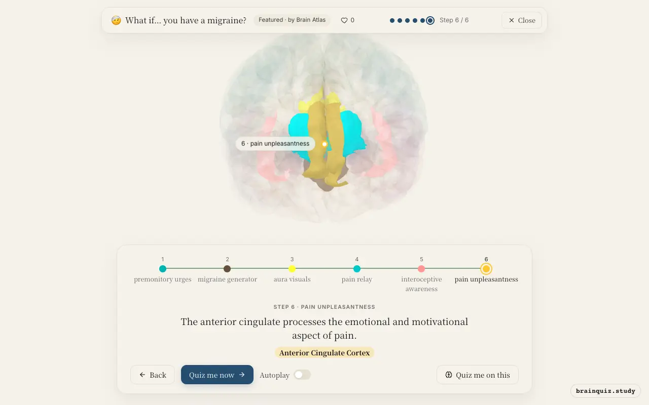
Watch it light up on the 3D brain
Brain regions that light up
| Order | Region | What it does here |
|---|---|---|
| 1 | Hypothalamus | The hypothalamus shifts activity, causing cravings, mood changes, or yawning before pain. |
| 2 | Brainstem | Brainstem nuclei become active, potentially triggering the migraine attack. |
| 3 | Visual Cortex | A wave of depressed activity spreads across the visual cortex, causing aura. |
| 4 | Thalamus | The thalamus relays trigeminal pain signals to the cortex. |
| 5 | Insula | The insula integrates pain and internal body signals, making you feel unwell. |
| 6 | Anterior Cingulate Cortex | The anterior cingulate processes the emotional and motivational aspect of pain. |
1. Hypothalamus: premonitory urges
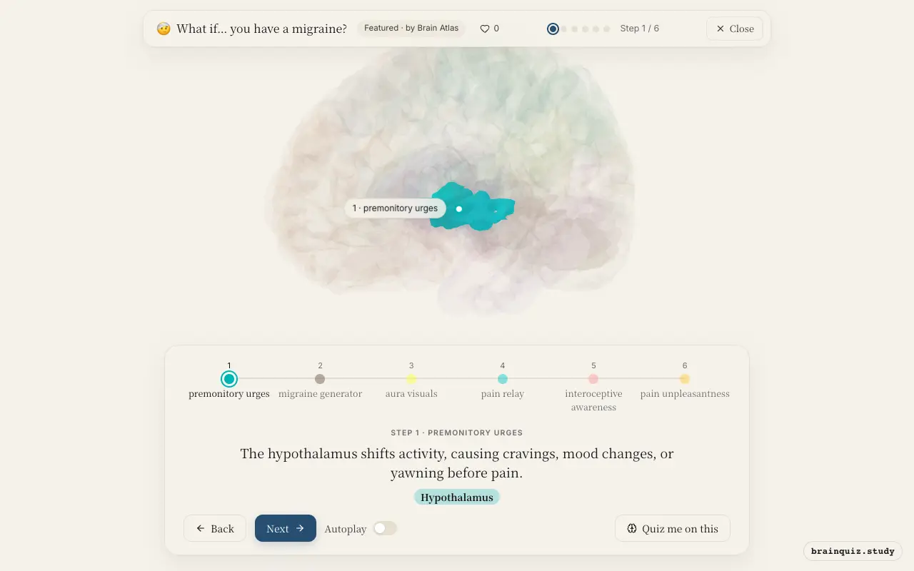
Changes in hypothalamic function are thought to initiate the premonitory phase, which can occur hours or days before headache. This region regulates homeostasis, circadian rhythms, and autonomic responses, linking internal states and external triggers to migraine onset. Its altered activity may explain symptoms like food cravings, mood swings, and yawning.
2. Brainstem: migraine generator
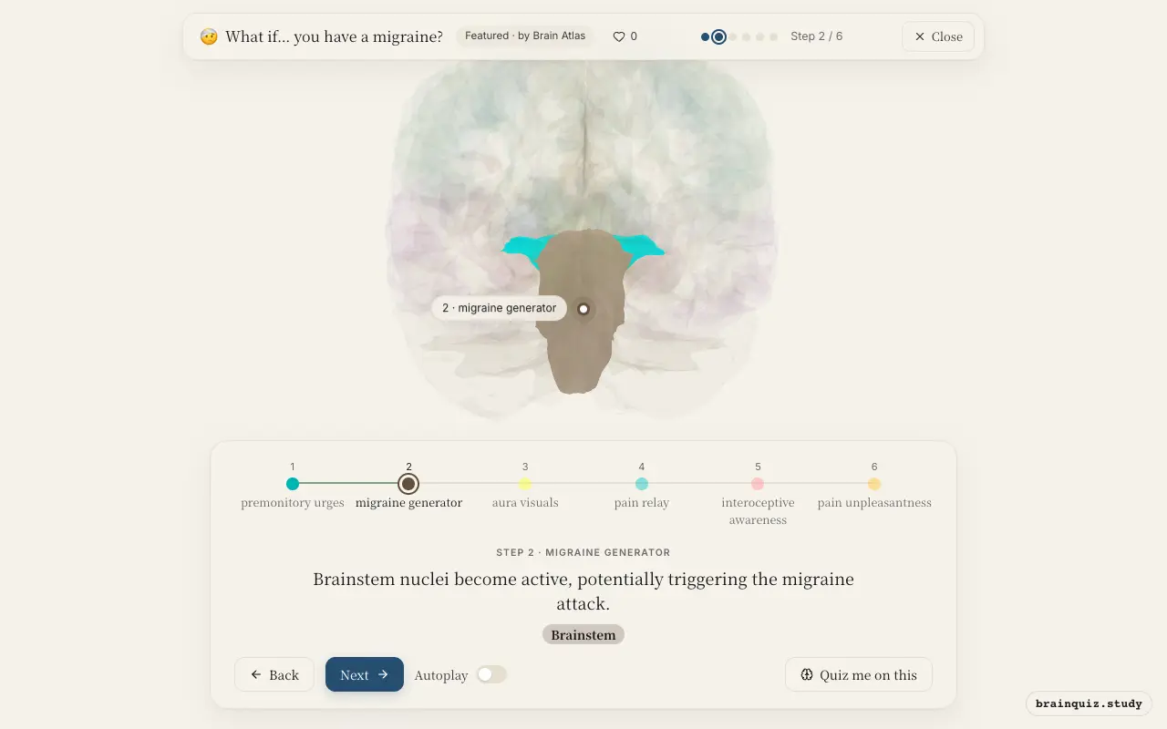
The brainstem, including the periaqueductal grey and trigeminal nucleus caudalis, is implicated in migraine generation. Abnormal activation of these regions may lower the threshold for pain signals and initiate the headache phase. Functional imaging studies suggest brainstem dysfunction is central to migraine pathophysiology.
3. Visual Cortex: aura visuals
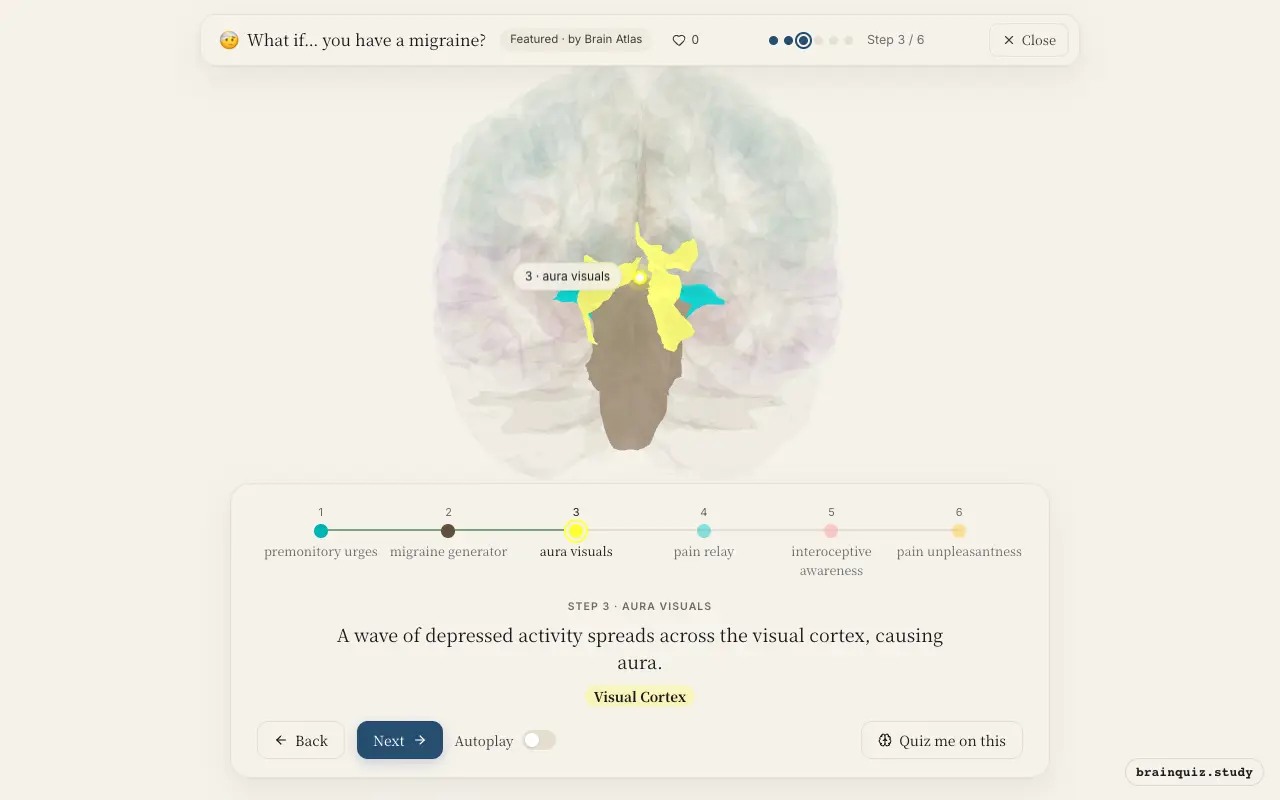
Cortical spreading depression is a slow wave of neuronal depolarization followed by suppression, often originating in the occipital lobe. This explains visual aura symptoms like scintillating scotomas or zigzag lines. It may also activate trigeminal afferents, linking aura to headache.
4. Thalamus: pain relay
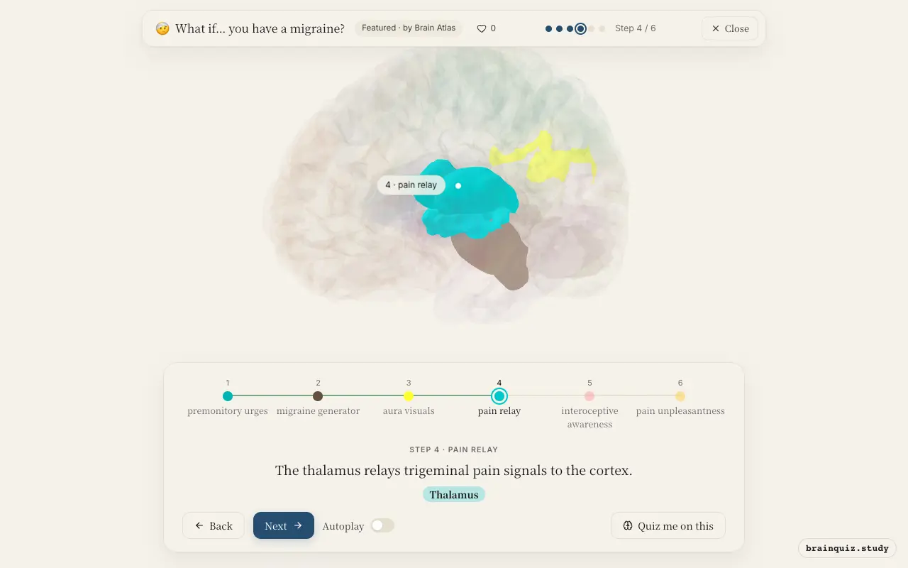
The thalamus acts as the main sensory relay station for pain signals. During migraine, it processes and forwards nociceptive inputs from the trigeminovascular system to cortical areas. Thalamic sensitization is thought to contribute to allodynia and the spread of pain beyond the original site.
5. Insula: interoceptive awareness
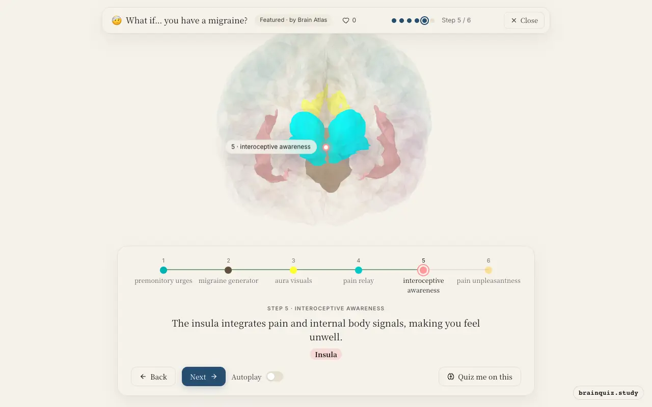
The insula is key for interoception, mapping internal bodily states. In migraine, it is consistently activated during pain, contributing to the sense of discomfort, nausea, and other autonomic symptoms. Insular activity may correlate with pain intensity, but not the overall migraine experience.
6. Anterior Cingulate Cortex: pain unpleasantness
The anterior cingulate cortex is part of the pain matrix, encoding the unpleasantness and the urge to escape pain. In migraine, its activation reflects the suffering component of headache. It may also be involved in the cognitive and affective changes.
Good to know
- Cortical spreading depression is the electrophysiological correlate of migraine aura.
- The trigeminovascular system, involving the trigeminal nerve and its central connections, is central to migraine headache.
- Migraine is a common neurological disorder, affecting over 10% of people worldwide.
Migraine vs Tension Headache
Migraine involves brainstem, cortex, and trigeminal system with throbbing pain, nausea, and sensitivity to light. Tension headache often involves pericranial muscle tenderness and stress, with dull bilateral pain and possible central sensitization. In tension headache, brainstem and thalamic activation is not consistently less pronounced.
Test yourself
Which region is thought to drive the premonitory phase of migraine?
What is the neural event responsible for migraine aura?
Which structure is central to relaying headache pain to the cortex?
0 of 3 answered · 0 correct
Simplified: many regions work together at once, and this shows the main players in the order they act. For learning only, not medical advice.