What happens in your brain when you grow old?
Updated
As you age, your prefrontal cortex shrinks, slowing executive functions; your hippocampus loses volume, impairing memory; your cerebellum loses cells, affecting coordination; your substantia nigra makes less dopamine, affecting movement; and your corpus callosum thins, slowing communication between hemispheres.
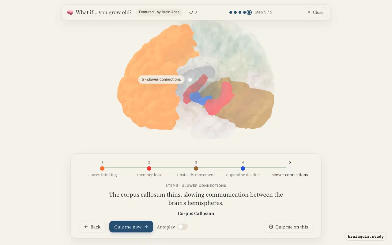
Watch it light up on the 3D brain
Brain regions that light up
| Order | Region | What it does here |
|---|---|---|
| 1 | Prefrontal Cortex | The prefrontal cortex shrinks with age, reducing its ability to manage attention and working memory. |
| 2 | Hippocampus | The hippocampus loses volume, impairing the formation of new memories. |
| 3 | Cerebellum | The cerebellum loses Purkinje cells, affecting balance and coordination. |
| 4 | Substantia Nigra | Dopamine-producing neurons in the substantia nigra decline, affecting movement and cognition. |
| 5 | Corpus Callosum | The corpus callosum thins, slowing communication between the brain's hemispheres. |
1. Prefrontal Cortex: slower thinking
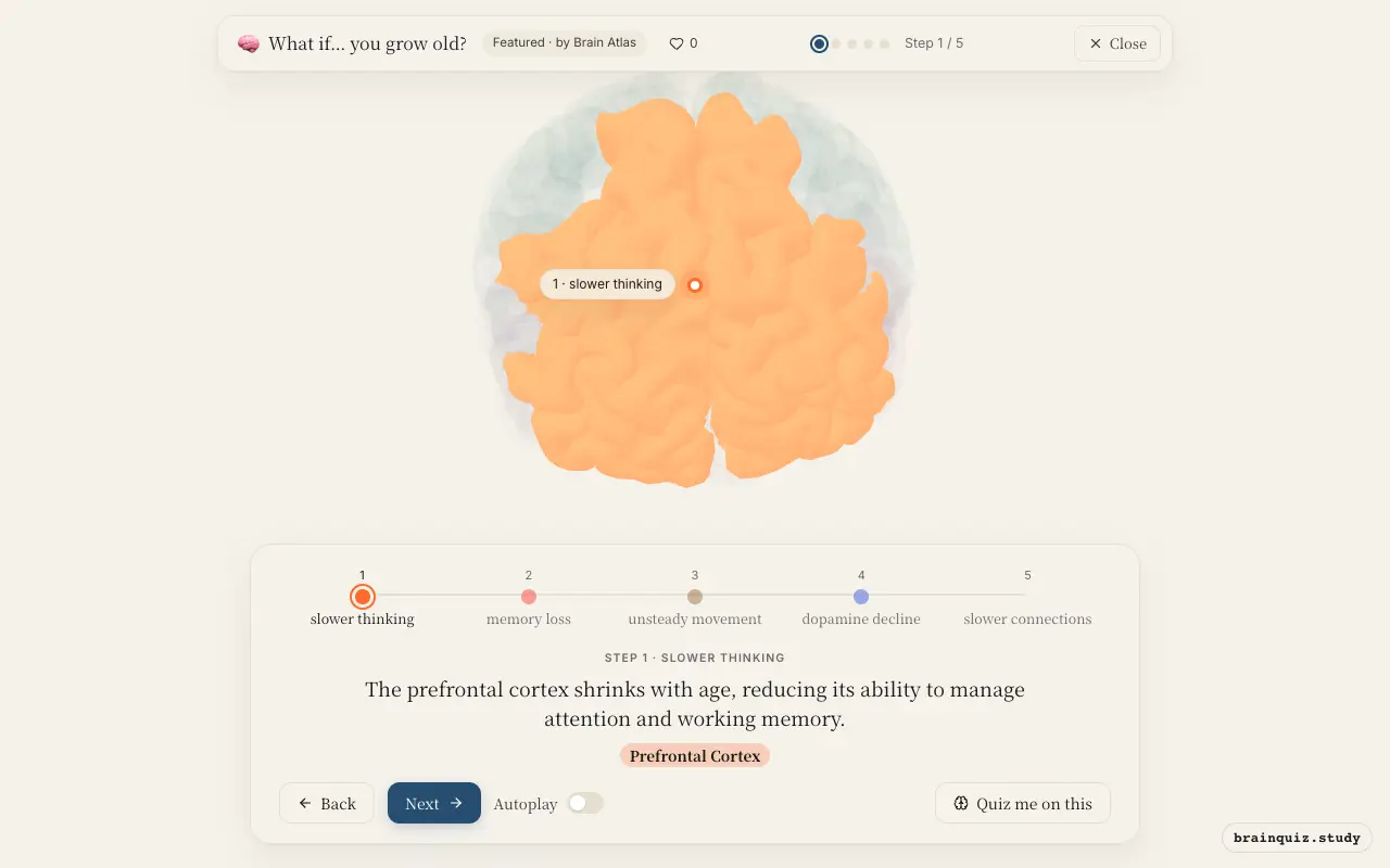
The prefrontal cortex, critical for executive functions like planning, decision-making, and working memory, shows age-related loss of gray matter and reduced synaptic density. This leads to slower processing, difficulty multitasking, and decreased mental flexibility. These changes are thought to contribute to the 'senior moments' often reported by older adults.
2. Hippocampus: memory loss
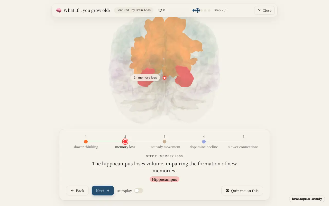
The hippocampus is essential for converting short-term memories into long-term storage. With age, it shrinks, and some evidence suggests reduced neurogenesis, but this is debated. This decline is linked to more difficulty learning new information and recalling recent events, though long-term memories often remain intact.
3. Cerebellum: unsteady movement
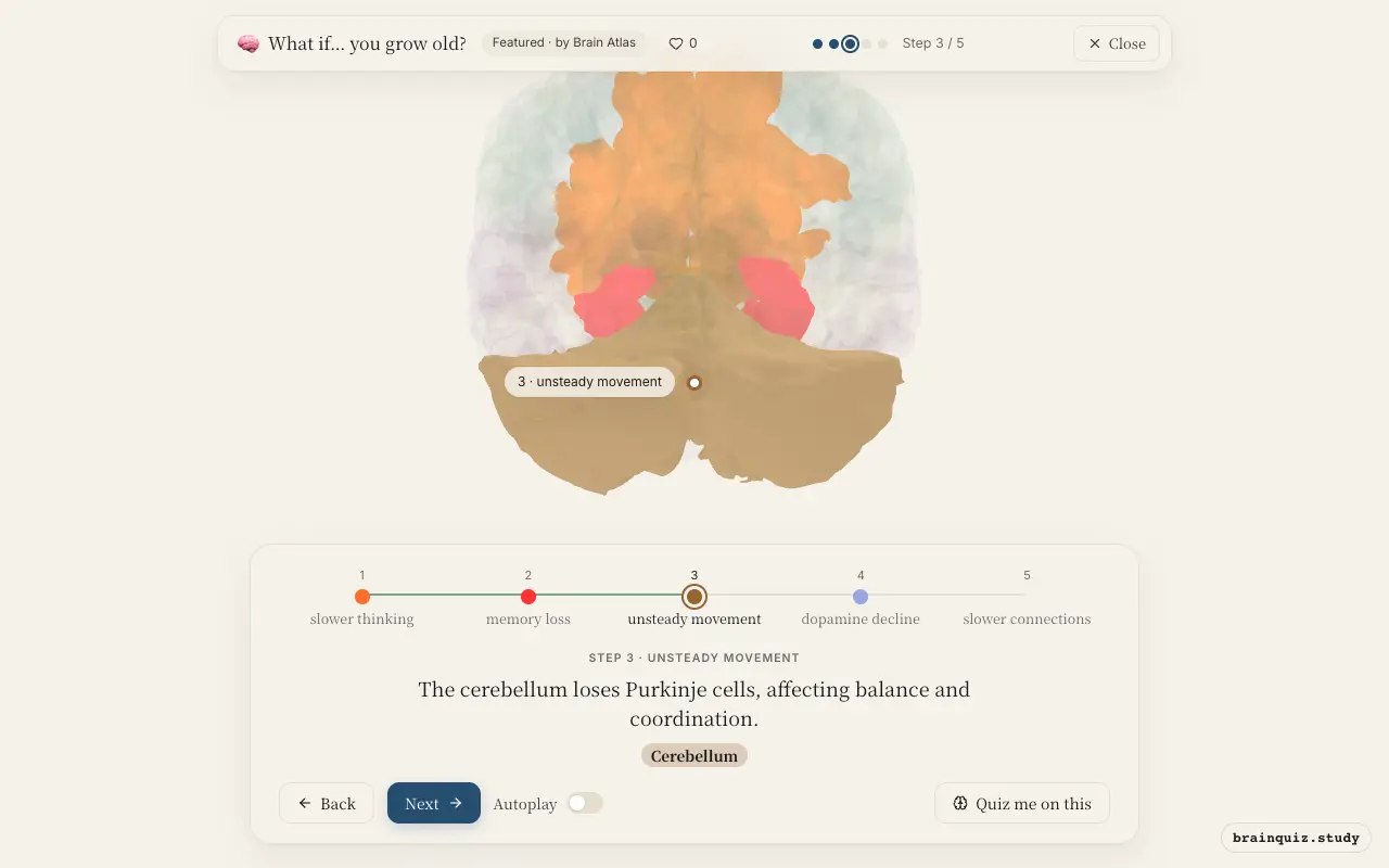
The cerebellum coordinates smooth movements, balance, and motor learning. Aging leads to a loss of Purkinje cells and reduced cerebellar volume, which contributes to slower gait, increased sway, and poorer fine motor skills. These changes are subtle in healthy aging but can increase fall risk.
4. Substantia Nigra: dopamine decline
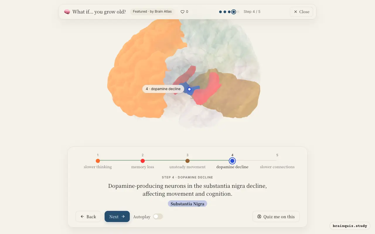
The substantia nigra supplies dopamine to the basal ganglia, which is crucial for initiating and controlling movement. Normal aging sees a gradual loss of these neurons, leading to slower movements and reduced motivation. While not as severe as in Parkinson's disease, this decline contributes to age-related slowing.
5. Corpus Callosum: slower connections
The corpus callosum is the main white matter tract connecting the left and right hemispheres. With age, its fibers lose myelin integrity, reducing the speed of interhemispheric communication. This may contribute to slower processing and less efficient coordination of tasks that require both hemispheres.
Good to know
- Some studies suggest the hippocampus may produce new neurons in adulthood, but this is debated, and such neurogenesis likely declines with age.
- White matter volume peaks around middle age and then declines, affecting processing speed.
- Normal aging is associated with a decline in dopamine receptors and transporters, which affects both motor and cognitive functions.
Normal aging vs Alzheimer's disease
In normal aging, brain changes are mild and gradual, with slight shrinkage and slowed communication. In Alzheimer's, there is severe atrophy, especially in the hippocampus and cortex, along with amyloid plaques and neurofibrillary tangles, causing profound memory loss and cognitive decline.
Test yourself
Which brain region is most associated with age-related memory decline?
What happens to the corpus callosum in normal aging?
Which neurotransmitter system declines with age, affecting both movement and cognition?
0 of 3 answered · 0 correct
Simplified: many regions work together at once, and this shows the main players in the order they act. For learning only, not medical advice.