What happens in your brain when you have a phobia?
Updated
In a phobia, the thalamus relays the feared stimulus to the amygdala, which triggers the hypothalamus for a rapid autonomic fear response; the insula registers bodily sensations, while the prefrontal cortex tries to regulate the reaction.
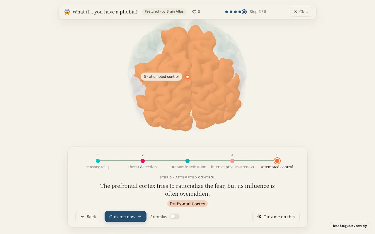
Watch it light up on the 3D brain
Brain regions that light up
| Order | Region | What it does here |
|---|---|---|
| 1 | Thalamus | The feared object or situation is sensed and sent to the thalamus for initial processing. |
| 2 | Amygdala | The amygdala instantly interprets the stimulus as extremely dangerous, sounding the alarm. |
| 3 | Hypothalamus | The hypothalamus triggers the fight-or-flight response through the autonomic nervous system. |
| 4 | Insula | The insula senses the racing heart and other bodily changes, amplifying the feeling of fear. |
| 5 | Prefrontal Cortex | The prefrontal cortex tries to rationalize the fear, but its influence is often overridden. |
1. Thalamus: sensory relay
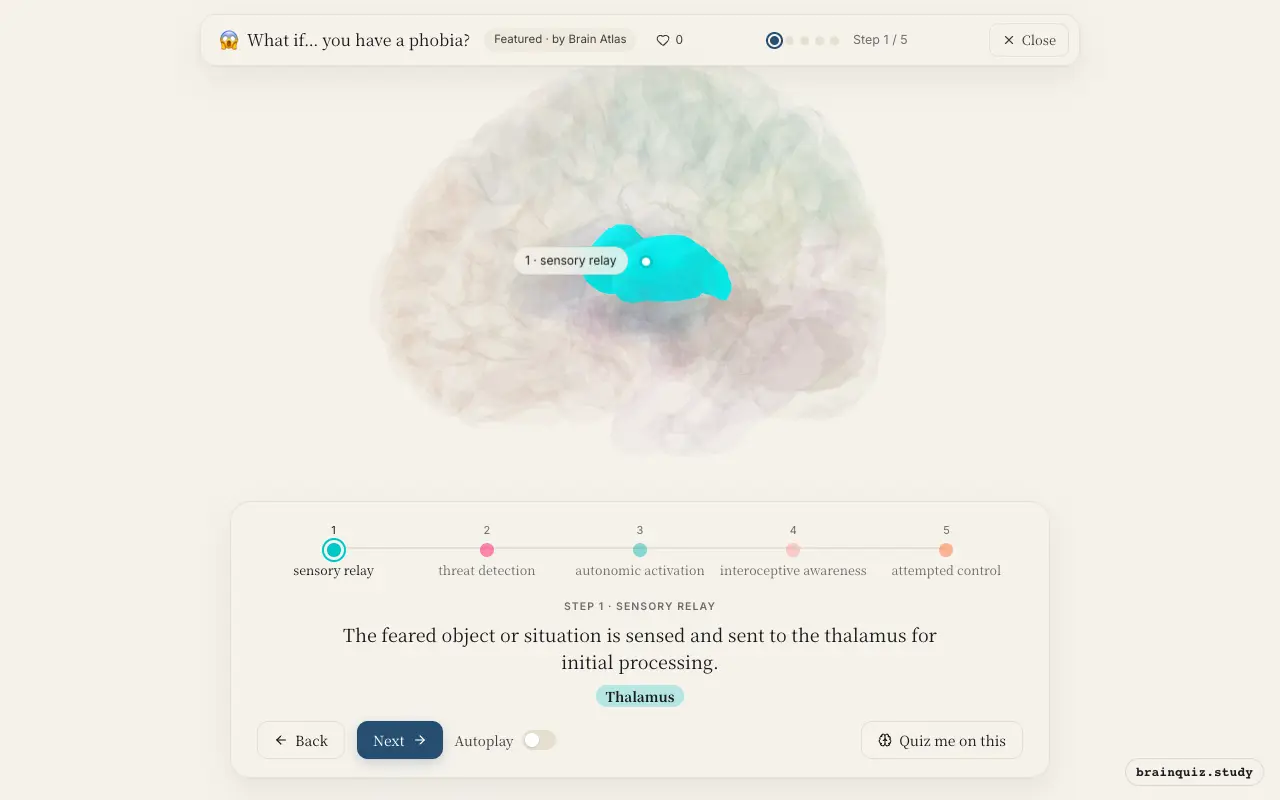
The thalamus acts as the brain's relay station, receiving sensory input from eyes, ears, or skin. In a phobia, it quickly routes the perceived threat to the amygdala, bypassing detailed cortical analysis to enable a fast response. This rapid pathway is evolutionarily conserved.
2. Amygdala: threat detection
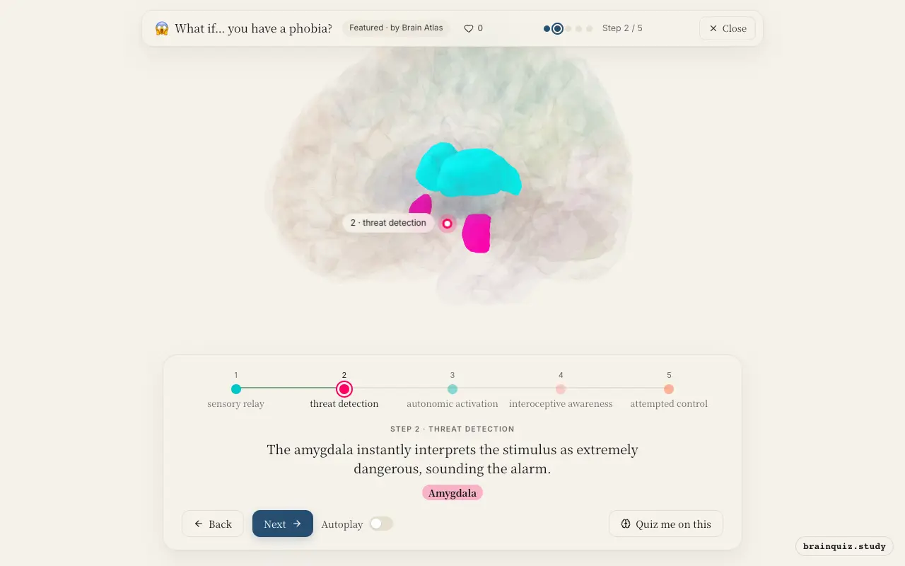
The amygdala is the brain's fear center. In phobia, it overreacts to a specific stimulus, triggering a cascade of fear responses even when the actual danger is minimal. This hyperactivity is thought to be a core feature of phobic disorders.
3. Hypothalamus: autonomic activation
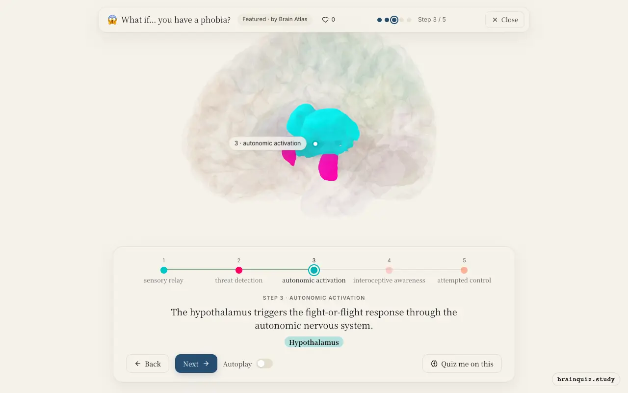
The hypothalamus activates the sympathetic nervous system, causing rapid heartbeat, sweating, and increased breathing. It also initiates the release of stress hormones like adrenaline. These bodily changes prepare the body to confront or escape the perceived threat.
4. Insula: interoceptive awareness
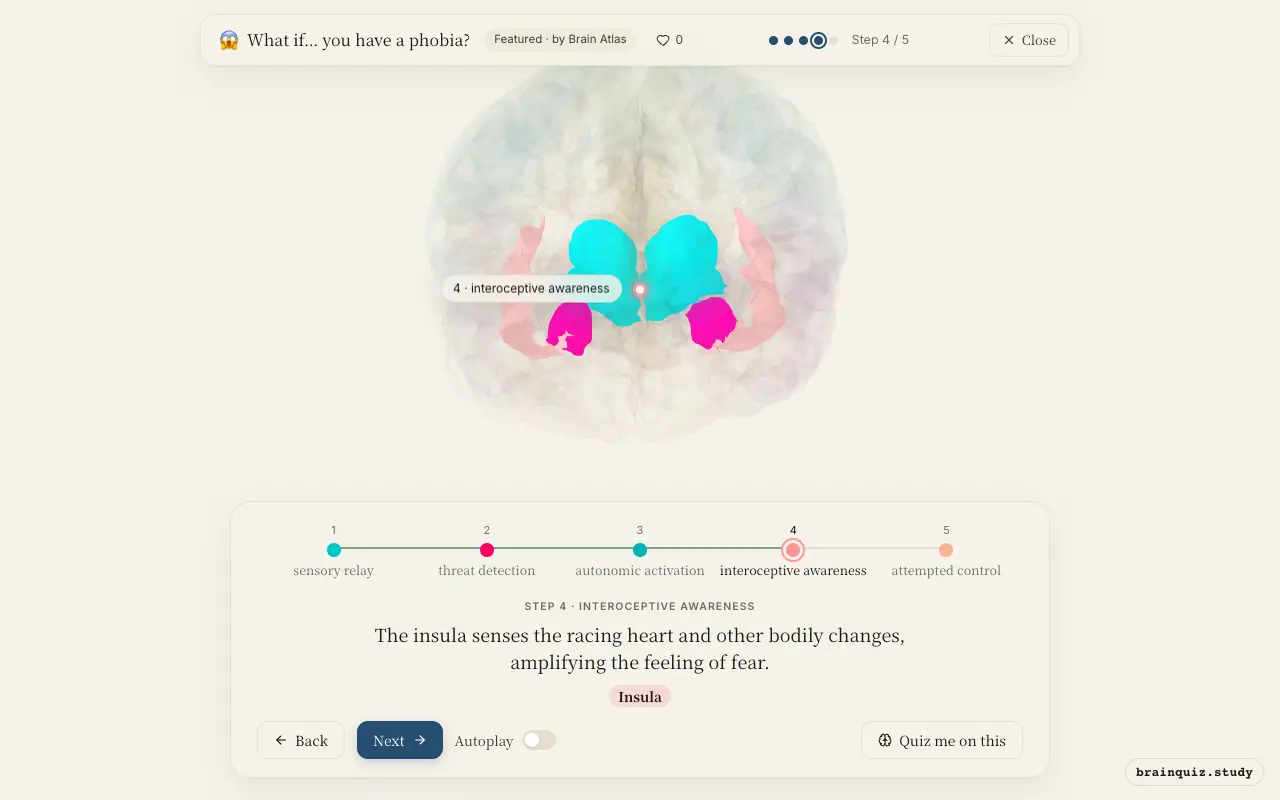
The insula maps internal bodily states, making you consciously aware of symptoms like a pounding heart or shortness of breath. In a phobia, this interoceptive feedback can intensify anxiety, creating a vicious cycle. Studies suggest the insula is more active during phobic reactions.
5. Prefrontal Cortex: attempted control
The prefrontal cortex, especially its medial and ventrolateral parts, normally regulates emotions by reappraising threats. In phobia, this top-down control is weakened, allowing the amygdala to dominate. Therapy aims to strengthen prefrontal regulation to reduce phobic responses.
Good to know
- The amygdala can trigger a fear response before the cortex fully processes the stimulus, enabling split-second reactions.
- Phobias are classified as anxiety disorders and often develop through classical conditioning or observational learning.
- Exposure therapy helps phobias by repeatedly activating the fear circuit in a safe context, gradually reducing the amygdala's response.
Phobia vs normal fear
In normal fear, the amygdala activates appropriately to a real threat and the prefrontal cortex regulates it once safety is confirmed. In a phobia, the amygdala fires excessively and the prefrontal cortex fails to dampen it, leading to persistent anxiety even when the feared object is harmless or absent.
Test yourself
Which brain region is primarily responsible for detecting the feared stimulus and initiating the fear response in a phobia?
What role does the prefrontal cortex typically play in a phobia?
Which pathway allows the amygdala to respond to a phobic stimulus before conscious awareness?
0 of 3 answered · 0 correct
Simplified: many regions work together at once, and this shows the main players in the order they act. For learning only, not medical advice.