What happens in your brain when you have ADHD?
Updated
ADHD involves underactivity in the prefrontal cortex (poor executive control), altered dopamine in the caudate and putamen (impaired reward and motor regulation), and cerebellar differences affecting timing and attention.
Watch it light up on the 3D brain
Brain regions that light up
| Order | Region | What it does here |
|---|---|---|
| 1 | Prefrontal Cortex | The prefrontal cortex is underactive, weakening executive control over attention and impulses. |
| 2 | Caudate Nucleus | The caudate nucleus shows altered dopamine transmission, impairing goal-directed behavior and reward learning. |
| 3 | Putamen | The putamen's role in motor control is disrupted, contributing to hyperactivity and restlessness. |
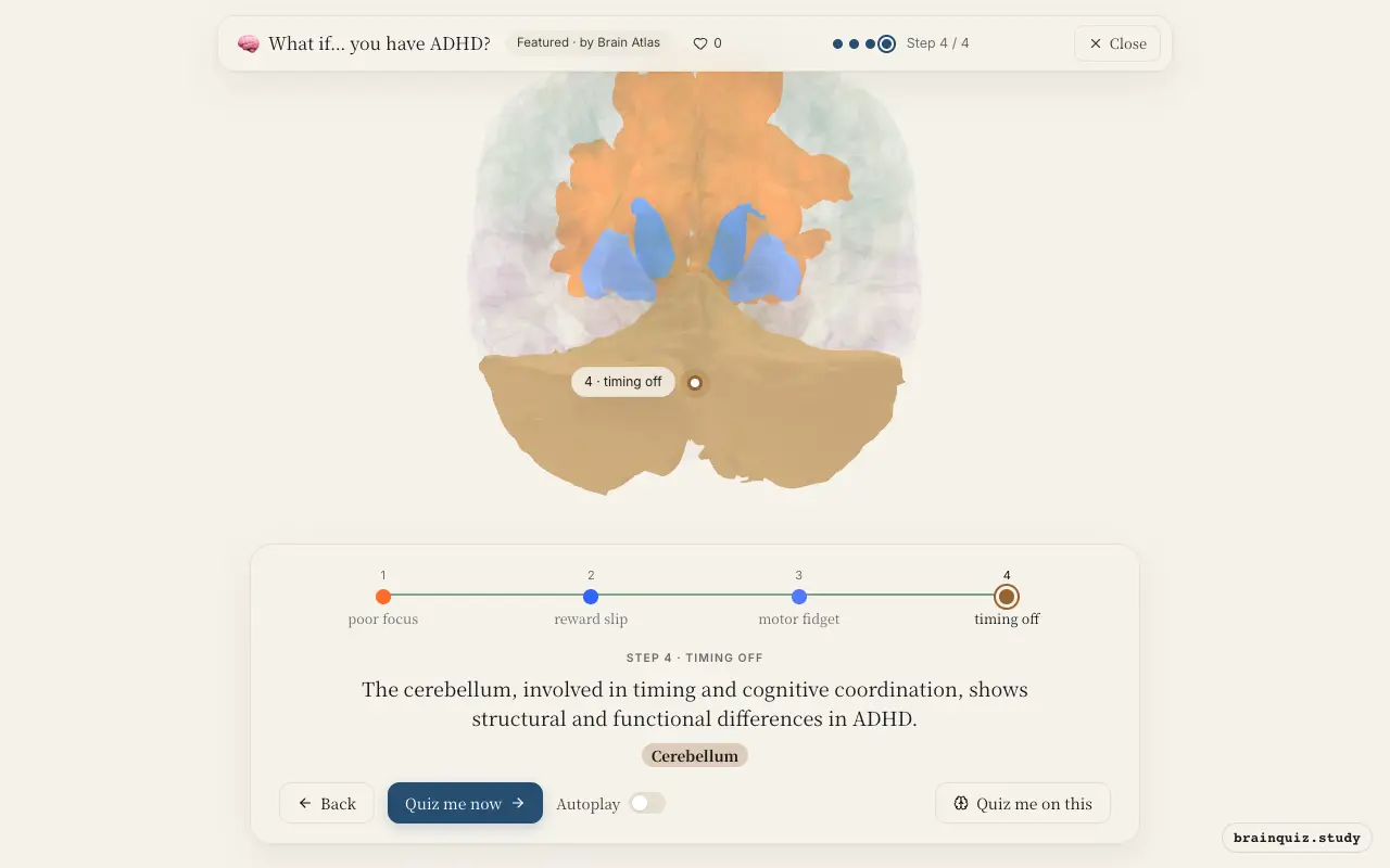
| 4 | Cerebellum | The cerebellum, involved in timing and cognitive coordination, shows structural and functional differences in ADHD. |
1. Prefrontal Cortex: poor focus
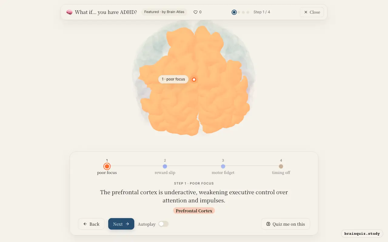
In ADHD, neuroimaging studies show reduced activation in the prefrontal cortex during tasks requiring sustained attention and response inhibition. This region normally supports working memory, planning, and self-regulation, so its underactivity leads to distractibility and impulsive actions. Dopamine and norepinephrine signaling here is often dysregulated.
2. Caudate Nucleus: reward slip
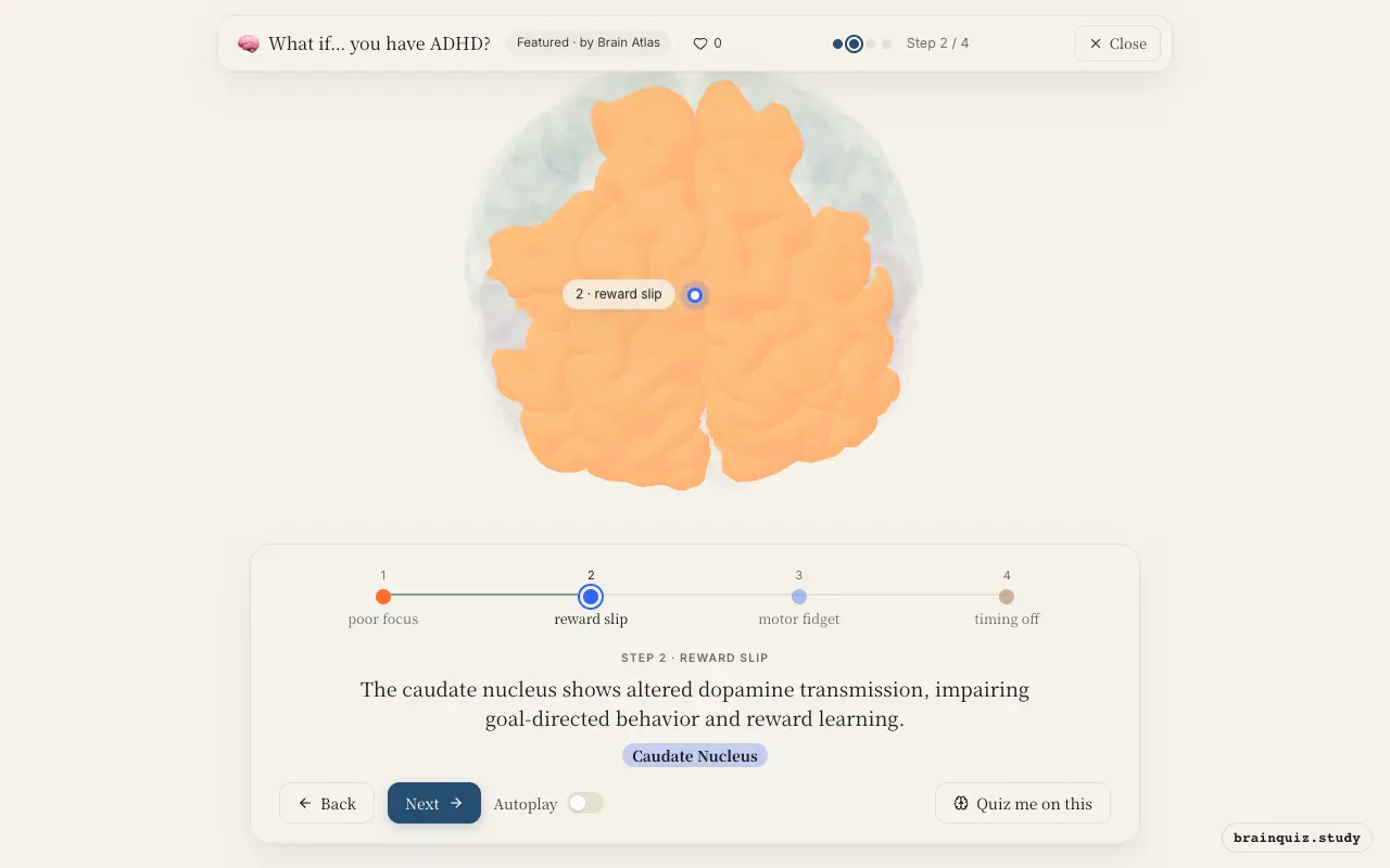
The caudate is part of the striatum and is critical for linking actions to outcomes and maintaining motivation. In ADHD, lower dopamine levels or receptor availability in the caudate are linked to difficulties with delayed gratification and staying on task. Stimulant medications increase dopamine here to improve symptoms.
3. Putamen: motor fidget
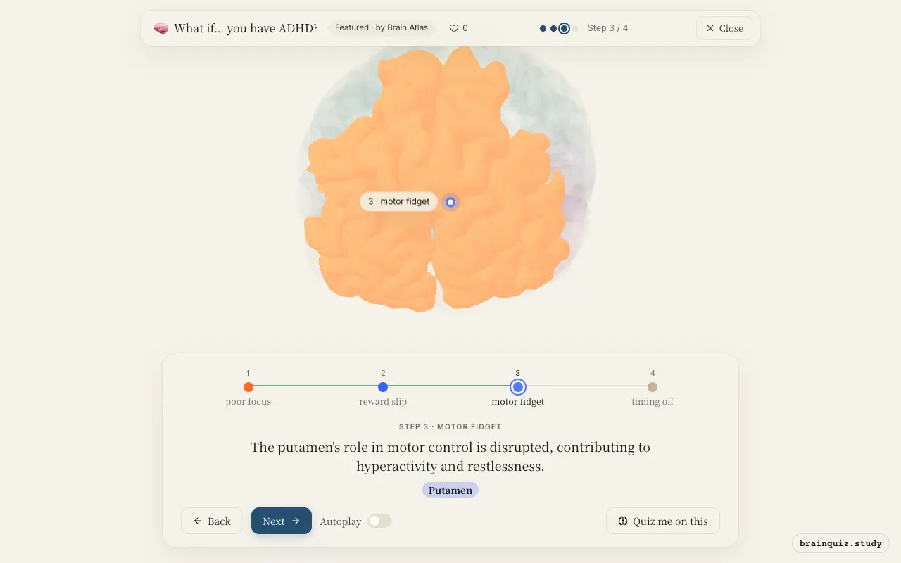
The putamen helps regulate habitual movements and inhibits unwanted motor activity. In ADHD, differences in putamen volume or activity are associated with excessive fidgeting, squirming, and difficulty sitting still. This is part of the broader fronto-striatal circuit dysfunction.
4. Cerebellum: timing off
Although traditionally linked to motor coordination, the cerebellum also contributes to attention and temporal processing. Studies have found smaller cerebellar volumes and altered connectivity in individuals with ADHD, which may underlie problems with time estimation and sequencing tasks.
Good to know
- ADHD is highly heritable, with family and twin studies showing strong genetic influences.
- Meta-analyses of brain imaging find reduced gray matter volume in the basal ganglia and prefrontal cortex in individuals with ADHD.
- Stimulant medications used to treat ADHD primarily act by increasing dopamine and norepinephrine availability in the brain.
ADHD vs. everyday distraction
Everyday distraction is transient and tied to fatigue or boredom, with normal prefrontal and striatal activity. In ADHD, the underactivity in these circuits is chronic and pervasive, affecting multiple settings. Hyperfocus in ADHD, paradoxically, reflects dysregulated attention rather than strong executive control.
Test yourself
Which brain region is most directly associated with executive dysfunction in ADHD?
What neurotransmitter abnormality is central to ADHD?
Which basal ganglia structure is involved in reward learning and goal-directed behavior, and shows altered function in ADHD?
0 of 3 answered · 0 correct
Simplified: many regions work together at once, and this shows the main players in the order they act. For learning only, not medical advice.