What happens in your brain when you have PTSD flashbacks?
Updated
PTSD flashbacks involve an overactive amygdala sounding a false alarm, a hippocampus that fails to contextualize the memory as past, and an underactive prefrontal cortex unable to inhibit the fear response, leading to vivid sensory re-experiencing and bodily panic via the insula.
Watch it light up on the 3D brain
Brain regions that light up
| Order | Region | What it does here |
|---|---|---|
| 1 | Amygdala | The amygdala detects a threat trigger and fires an immediate fear response. |
| 2 | Hippocampus | The hippocampus fails to tag the memory as past, making it feel present. |
| 3 | Prefrontal Cortex | The prefrontal cortex, especially its medial parts, fails to inhibit the amygdala. |
| 4 | Visual Cortex | The visual cortex re-creates the traumatic scene as if it were real. |
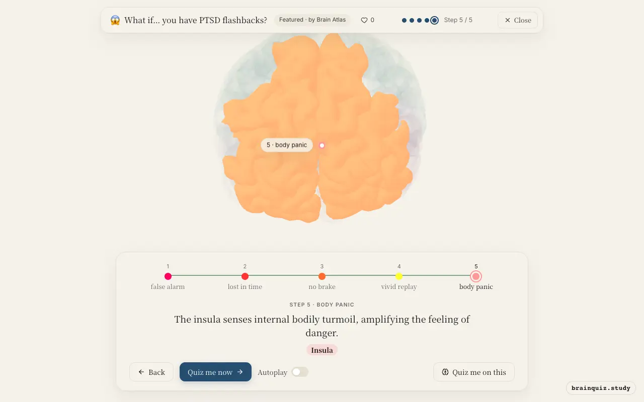
| 5 | Insula | The insula senses internal bodily turmoil, amplifying the feeling of danger. |
1. Amygdala: false alarm
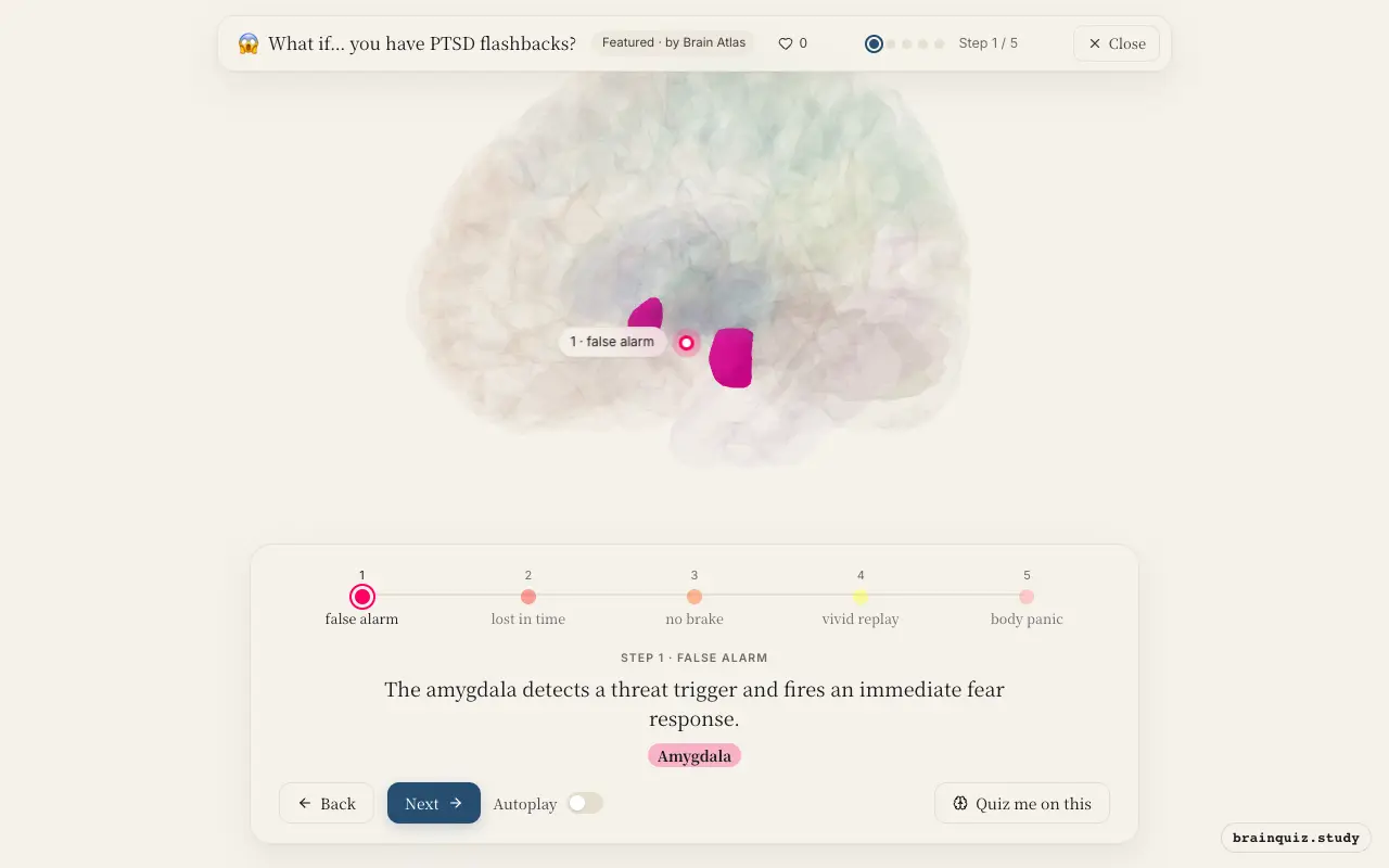
In PTSD, the amygdala is hyperresponsive to trauma-related cues, even safe stimuli resembling the original event can trigger a full-blown fear response. This rapid, bottom-up alarm precedes conscious thought, preparing the body for fight or flight and bypassing slower cortical evaluation.
2. Hippocampus: lost in time
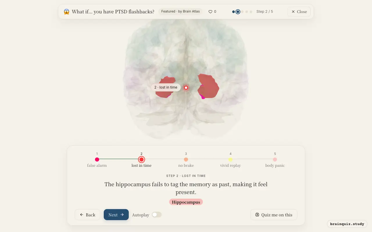
Normally, the hippocampus encodes the spatial and temporal context of memories, allowing you to recognize them as belonging to the past. In PTSD, hippocampal function is impaired, so the traumatic memory is retrieved without this context. As a result, the flashback feels like it is happening right now, not a recollection.
3. Prefrontal Cortex: no brake
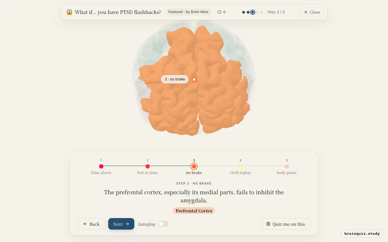
The medial prefrontal cortex normally exerts top-down control over the amygdala, dampening fear responses once a threat has passed. In PTSD, this region shows reduced activity and connectivity with the amygdala. Consequently, the amygdala remains unchecked, and the fear response escalates instead of being regulated.
4. Visual Cortex: vivid replay
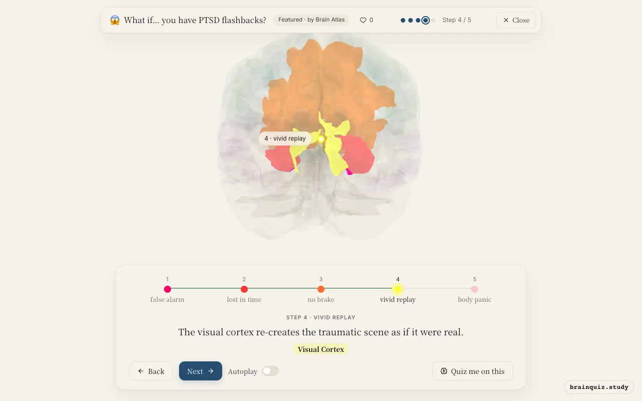
During a flashback, sensory cortices become highly active, driven by the amygdala's alarm. The visual cortex, in particular, generates vivid images of the traumatic event, contributing to the sense of re-experiencing. This is not a voluntary recall but an involuntary, perception-like activation.
5. Insula: body panic
The insula receives interoceptive signals from the body, such as heart rate, breathing, and gut sensations. In a flashback, it becomes hyperactive and interprets these physiological changes as evidence of immediate threat. This creates a feedback loop that intensifies the subjective sense of panic and fear.
Good to know
- Studies suggest that people with PTSD show increased amygdala activation and decreased prefrontal activation when exposed to trauma reminders.
- The hippocampus is thought to be smaller in many individuals with chronic PTSD, though causality is debated.
- Flashbacks can involve not only visual but also auditory, olfactory, and somatic re-experiencing.
Flashback vs Normal Recall
During normal recall, the hippocampus provides contextual details, and the prefrontal cortex keeps the amygdala in check, so the memory feels distant and safe. In a PTSD flashback, hippocampal context is missing, prefrontal inhibition fails, and the amygdala drives sensory re-experiencing, making the past feel present and threatening.
Test yourself
Which brain region is primarily responsible for the initial fear response in a PTSD flashback?
Why does a flashback feel like it is happening now rather than being remembered?
What role does the prefrontal cortex play in PTSD flashbacks?
0 of 3 answered · 0 correct
Simplified: many regions work together at once, and this shows the main players in the order they act. For learning only, not medical advice.