What happens in your brain when you lie awake with insomnia?
Updated
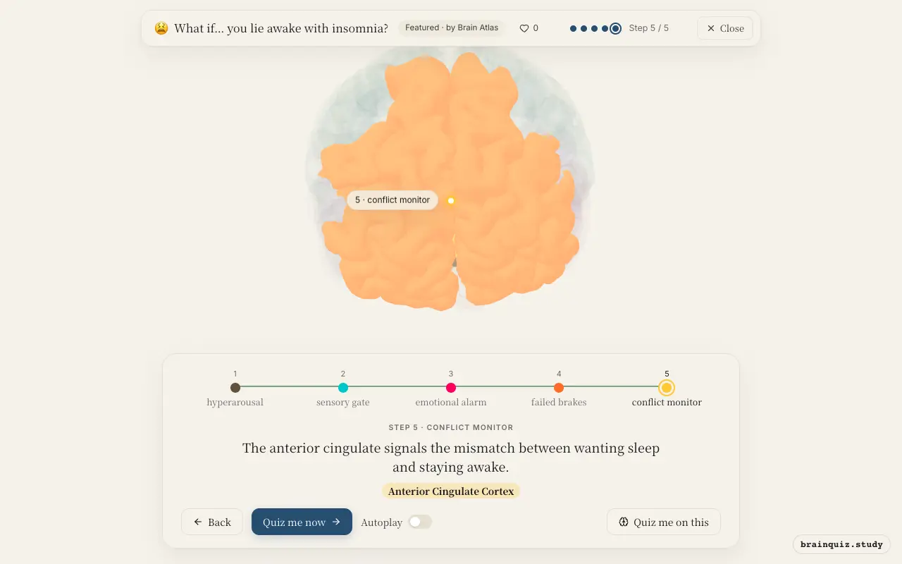
Your brainstem triggers hyperarousal, the thalamus keeps your cortex awake to sensory input, the amygdala amplifies emotional distress, the prefrontal cortex fails to stop rumination, and the anterior cingulate flags the sleep-wake conflict.
Watch it light up on the 3D brain
Brain regions that light up
| Order | Region | What it does here |
|---|---|---|
| 1 | Brainstem | The brainstem's arousal network stays active, preventing the transition to sleep. |
| 2 | Thalamus | The thalamus relays sensory information to the cortex, keeping you aware of your environment. |
| 3 | Amygdala | The amygdala detects emotional threats, amplifying distress and arousal. |
| 4 | Prefrontal Cortex | The prefrontal cortex tries to suppress racing thoughts but is overwhelmed. |
| 5 | Anterior Cingulate Cortex | The anterior cingulate signals the mismatch between wanting sleep and staying awake. |
1. Brainstem: hyperarousal
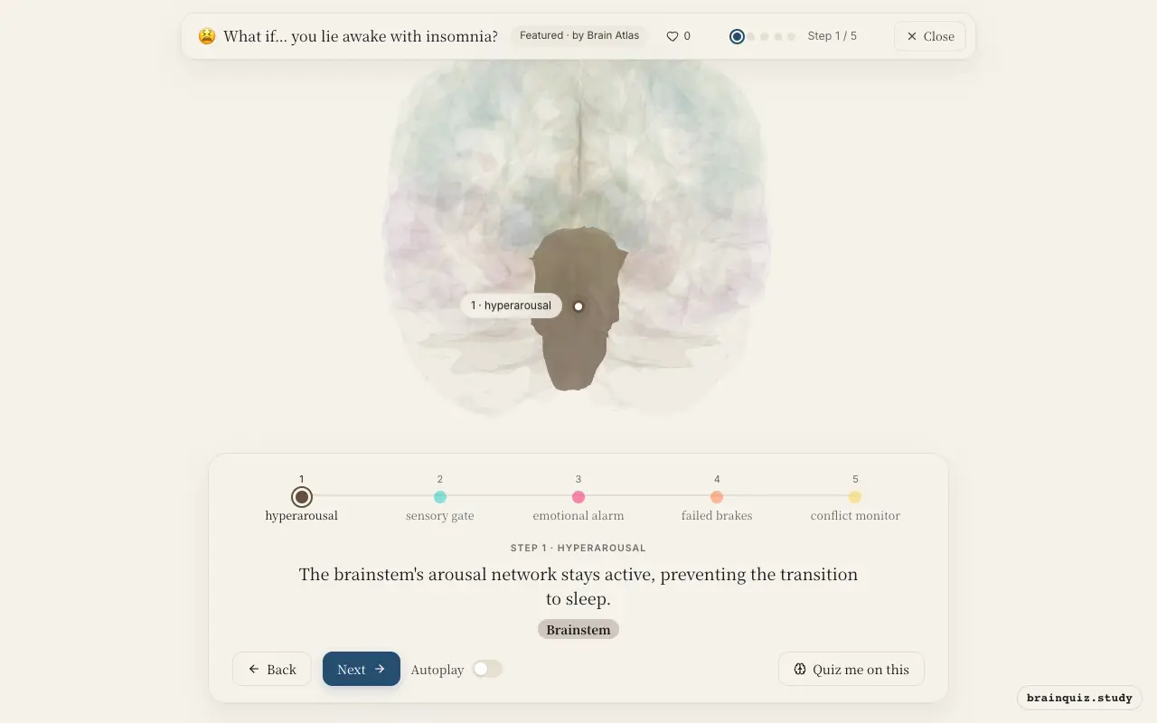
The brainstem contains the reticular activating system, which promotes wakefulness by sending excitatory signals to the thalamus and cortex. In insomnia, this system remains overactive due to stress or learned hypervigilance, keeping you alert instead of allowing the gradual decrease in firing that normally initiates sleep. This sustained arousal is a hallmark of the disorder.
2. Thalamus: sensory gate
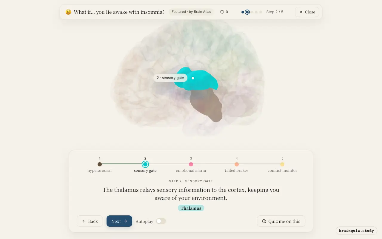
The thalamus acts as a relay station for sensory input, and during sleep onset it normally reduces transmission to allow cortical deactivation. In insomnia, the thalamus continues to pass sounds, touch, and other stimuli to the cortex, preventing the sensory disengagement needed for sleep. This heightened sensory processing contributes to the feeling of being 'on alert'.
3. Amygdala: emotional alarm
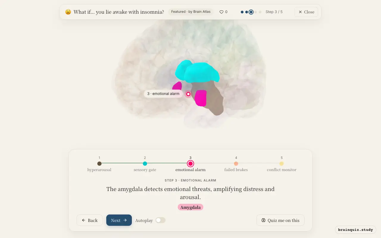
The amygdala is central to processing fear and emotional salience. During insomnia, it becomes hyperresponsive to negative thoughts or bodily sensations, triggering a stress response that further increases arousal. This emotional reactivity creates a feedback loop: worry activates the amygdala, which in turn fuels more worry and physiological arousal, making it harder to relax.
4. Prefrontal Cortex: failed brakes
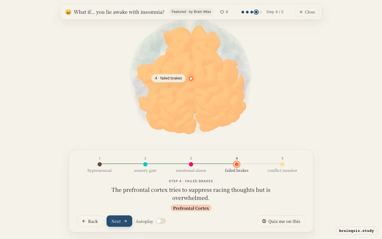
The prefrontal cortex normally exerts top-down control over the amygdala and other limbic areas, helping to regulate emotion and quiet the mind. In insomnia, this region may be less effective due to fatigue or stress, allowing intrusive thoughts and worries to persist. The inability to shift attention away from concerns keeps you mentally active and delays sleep.
5. Anterior Cingulate Cortex: conflict monitor
The anterior cingulate cortex detects when there is a conflict between your current state and your goal. When you lie awake wanting to sleep, it registers the discrepancy and generates a sense of frustration and urgency. This activation can increase arousal and negative affect, making you more aware of the problem and less able to let go.
Good to know
- Chronic insomnia is associated with elevated levels of cortisol and norepinephrine, reflecting a state of physiological hyperarousal.
- The default mode network, including the posterior cingulate cortex, shows increased connectivity during rumination, which is common in insomnia.
- Cognitive behavioral therapy for insomnia targets the reduction of pre-sleep cognitive arousal by training patients to redirect thoughts.
Insomnia vs. Normal Sleep Onset
During normal sleep onset, brainstem arousal systems and the thalamus gradually reduce activity, allowing cortical synchronization into slow waves. In insomnia, these systems remain active, the amygdala stays vigilant, and the prefrontal cortex fails to disengage from wakeful thought patterns, prolonging the transition to sleep.
Test yourself
Which brain region is primarily responsible for the hyperarousal state in insomnia?
What role does the anterior cingulate cortex play during insomnia?
How does the prefrontal cortex contribute to insomnia?
0 of 3 answered · 0 correct
Simplified: many regions work together at once, and this shows the main players in the order they act.