What happens in your brain when you look at a face?
Updated
Light hits the visual cortex for basic processing, then the fusiform gyrus identifies it as a face. The amygdala reads emotional expression, and the orbitofrontal cortex judges social meaning and response.
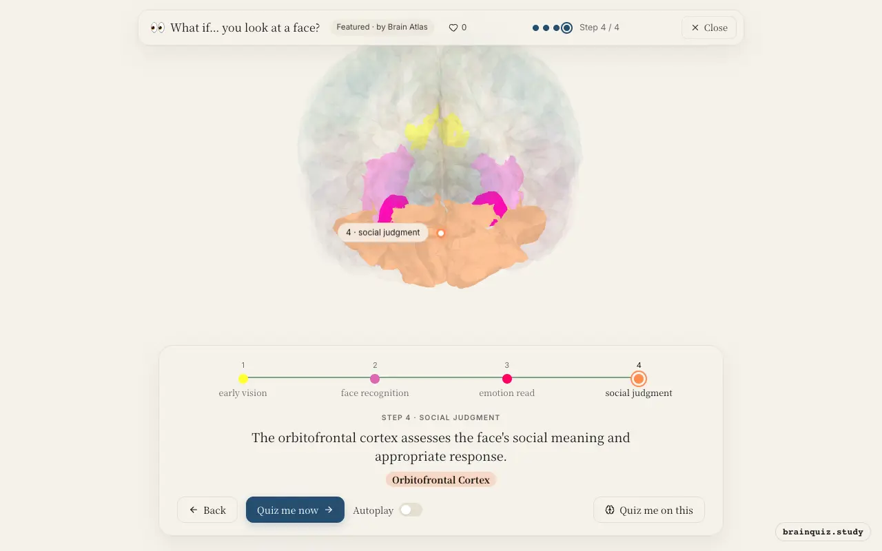
Watch it light up on the 3D brain
Brain regions that light up
| Order | Region | What it does here |
|---|---|---|
| 1 | Visual Cortex | The visual cortex detects edges, shapes, and contrast from the face. |
| 2 | Fusiform Gyrus | The fusiform gyrus matches the pattern to stored face templates. |
| 3 | Amygdala | The amygdala evaluates the facial expression for threat or social cues. |
| 4 | Orbitofrontal Cortex | The orbitofrontal cortex assesses the face's social meaning and appropriate response. |
1. Visual Cortex: early vision
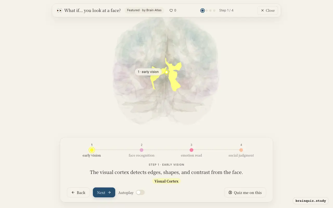
The primary visual cortex in the occipital lobe receives input from the eyes via the lateral geniculate nucleus of the thalamus. Its neurons detect edges, orientation, and spatial frequency, building a raw neural image of the face. This information is sent forward to the ventral visual stream for higher-level processing.
2. Fusiform Gyrus: face recognition
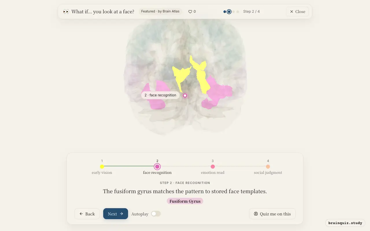
Located on the ventral surface of the temporal lobe, the fusiform face area is selectively responsive to faces. It integrates features like eyes, nose, and mouth into a holistic representation, allowing rapid identification of identity and species. Damage to this region can cause prosopagnosia, the inability to recognize faces.
3. Amygdala: emotion read
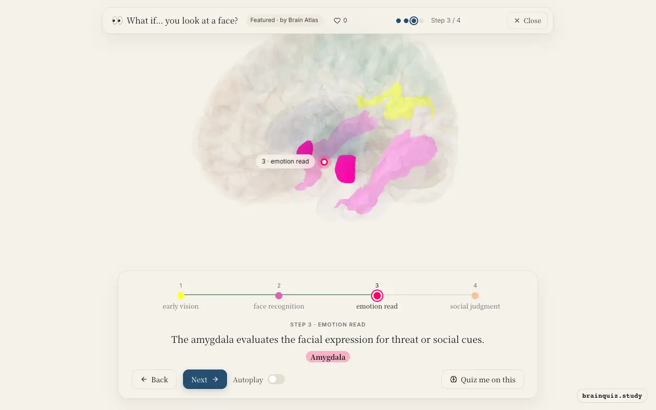
The amygdala receives fast, low-resolution input from the superior colliculus and pulvinar as well as detailed input from the fusiform gyrus. It responds strongly to fearful or angry faces, triggering arousal and autonomic responses. This rapid evaluation prepares the body for potential threat or social engagement.
4. Orbitofrontal Cortex: social judgment
The orbitofrontal cortex receives input from the amygdala, temporal cortex, and sensory association areas. It integrates the perceived emotion with past experience and current social context, assigning value and guiding behavior. This region is essential for adaptive social interactions, such as judging trustworthiness or attractiveness.
Good to know
- Face-selective neurons in the fusiform gyrus respond more strongly to upright than inverted faces.
- Some studies suggest the amygdala shows increased activity to fearful faces presented subliminally.
- Some patients with amygdala damage have difficulty recognizing fearful expressions.
Face vs Object Recognition
When looking at an object, the lateral occipital complex and general temporal cortex handle recognition, with less fusiform gyrus specialization. Faces additionally recruit the amygdala and orbitofrontal cortex for emotional and social evaluation, whereas objects typically do not trigger such limbic responses.
Test yourself
Which brain region is most specialized for face recognition?
What role does the amygdala play when seeing a face?
Damage to the fusiform gyrus can cause what condition?
0 of 3 answered · 0 correct
Simplified: many regions work together at once, and this shows the main players in the order they act.