What happens in your brain when you see an optical illusion?
Updated
Your visual cortex detects edges and patterns; the occipital cortex integrates motion and context; the parietal cortex applies spatial attention, while the prefrontal cortex sets expectations that bias perception.
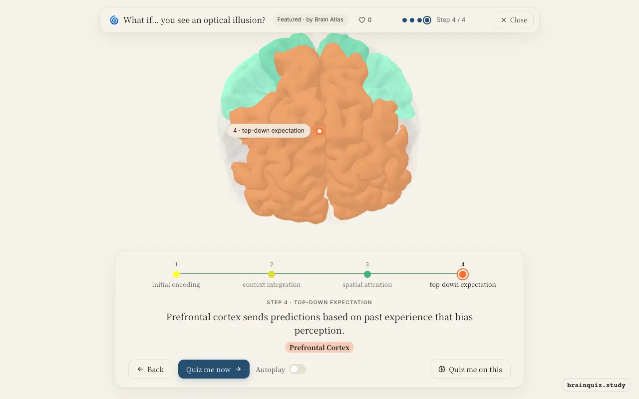
Watch it light up on the 3D brain
Brain regions that light up
| Order | Region | What it does here |
|---|---|---|
| 1 | Visual Cortex | Primary visual cortex processes edges, orientation, and contrast from the retinal image. |
| 2 | Occipital Cortex | Higher visual areas in occipital cortex integrate motion, color, and contextual information. |
| 3 | Parietal Cortex | Parietal cortex directs attention to relevant parts of the scene and applies spatial heuristics. |
| 4 | Prefrontal Cortex | Prefrontal cortex sends predictions based on past experience that bias perception. |
1. Visual Cortex: initial encoding
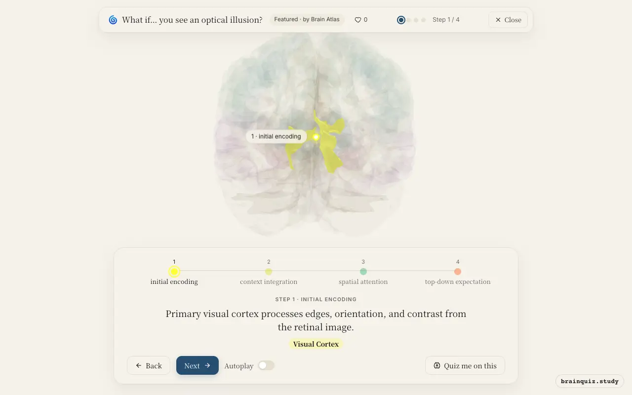
The visual cortex receives input from the thalamus and extracts fundamental features such as lines, angles, and luminance differences. Illusions often exploit these low-level processes, such as the simultaneous contrast effect where identical gray patches appear different due to surrounding context. This stage establishes the raw material for higher-level interpretation.
2. Occipital Cortex: context integration
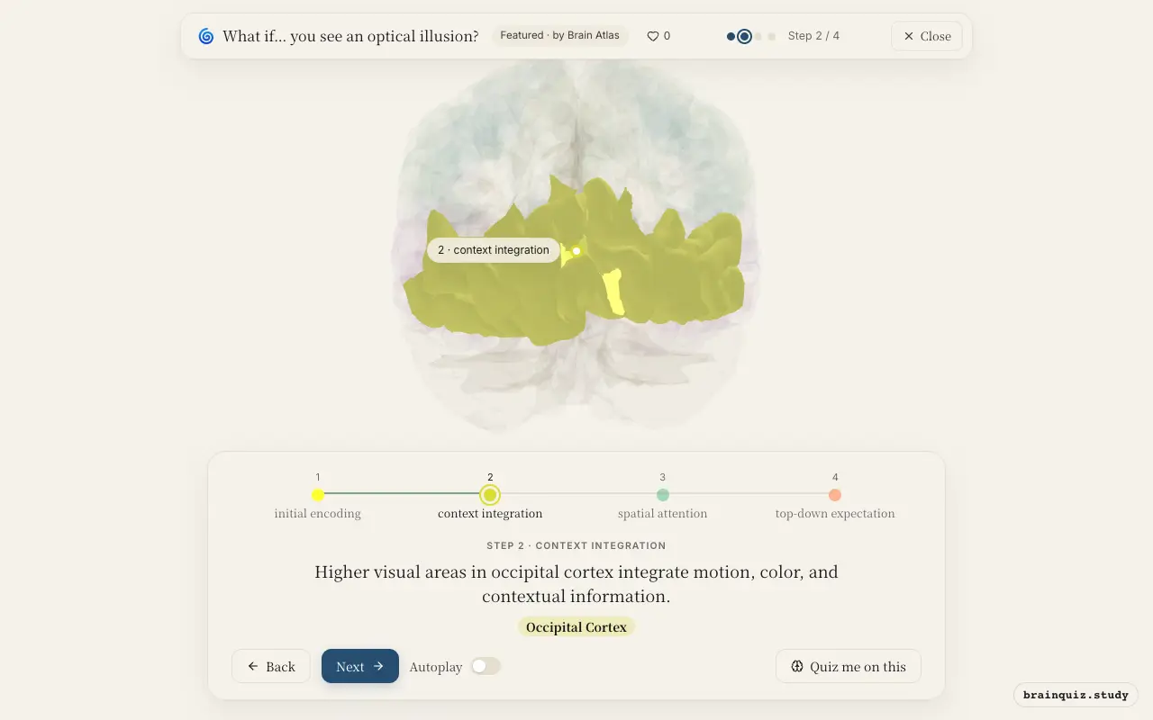
The occipital cortex includes regions like V2, V4, and MT that combine features into coherent percepts and incorporate surrounding information. For example, in the Ebbinghaus illusion, neurons here respond to the context of surrounding circles, altering perceived size. This integration often overrides the raw size information from the primary visual cortex.
3. Parietal Cortex: spatial attention
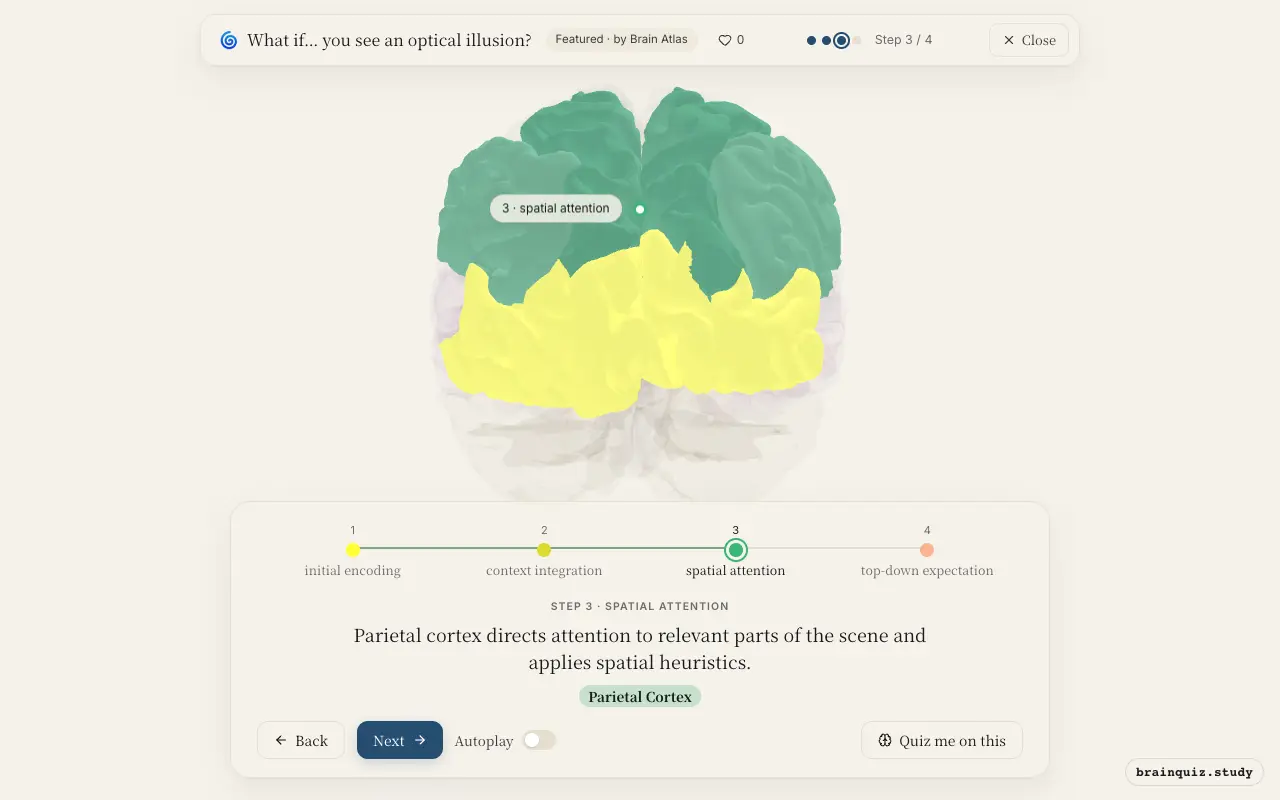
The parietal cortex is crucial for spatial attention and representing object locations. In illusions like the Müller-Lyer, it may activate when processing the arrowheads as depth cues, though this explanation is contested. Top-down signals from parietal areas modulate early visual responses, shaping the final illusion.
4. Prefrontal Cortex: top-down expectation
The prefrontal cortex holds expectations about the world, such as the assumption that objects have consistent size and shape. These priors are fed back to visual areas, creating the illusion when sensory evidence conflicts. In ambiguous figures, prefrontal activity increases as the brain resolves competing interpretations.
Good to know
- Optical illusions are not errors but byproducts of the brain's efficient use of context and prior experience to interpret ambiguous stimuli.
- The visual system has separate pathways for object identity (ventral stream, including occipitotemporal cortex) and spatial location (dorsal stream, including parietal cortex), which can disagree during illusions.
Illusion vs Hallucination
An optical illusion is a misinterpretation of a real external stimulus, engaging normal visual pathways with strong top-down modulation from prefrontal and parietal cortex. A hallucination is a perception without external stimulus, typically arising from abnormal spontaneous activity in sensory cortex or dysregulated dopamine. While both alter perception, illusions are shared and predictable, whereas hallucinations are idiosyncratic and often pathological.
Test yourself
Which brain region is the first cortical area to process visual input from the eyes?
Which brain region is most associated with applying prior knowledge and expectations to resolve an optical illusion?
In the Ebbinghaus illusion, where a central circle appears larger when surrounded by smaller circles, which brain region processes the contextual size information?
0 of 3 answered · 0 correct
Simplified: many regions work together at once, and this shows the main players in the order they act.