What happens in your brain when you meditate?
Updated
Meditation engages the prefrontal cortex for attention, anterior cingulate for monitoring, insula for body awareness, while posterior cingulate quiets and amygdala reactivity decreases.
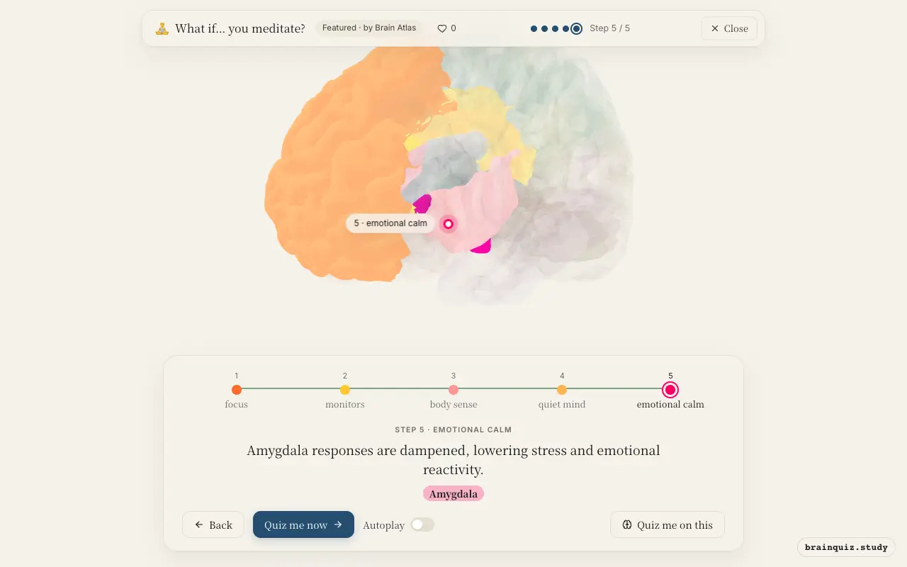
Watch it light up on the 3D brain
Brain regions that light up
| Order | Region | What it does here |
|---|---|---|
| 1 | Prefrontal Cortex | Prefrontal cortex sustains attention on the breath or chosen object. |
| 2 | Anterior Cingulate Cortex | Anterior cingulate detects mind-wandering and errors in focus. |
| 3 | Insula | Insula heightens interoceptive awareness of breathing and sensations. |
| 4 | Posterior Cingulate Cortex | Posterior cingulate decreases activity, reducing mind-wandering and self-referential thought. |
| 5 | Amygdala | Amygdala responses are dampened, lowering stress and emotional reactivity. |
1. Prefrontal Cortex: focus
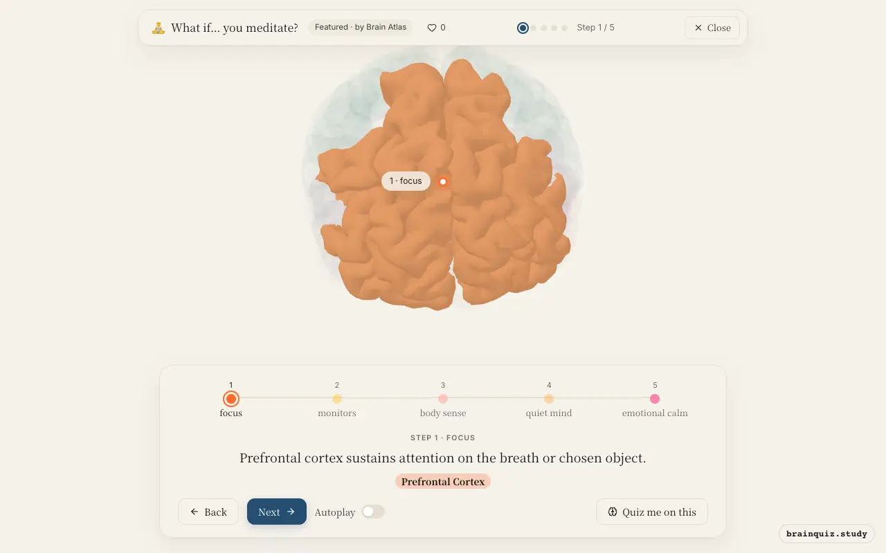
The prefrontal cortex, especially dorsolateral regions, is critical for maintaining goal-directed attention. During meditation, it keeps the mind anchored to the present moment, resisting the pull of distractions. This executive control is strengthened with regular practice, as studies suggest increased activity in this area.
2. Anterior Cingulate Cortex: monitors
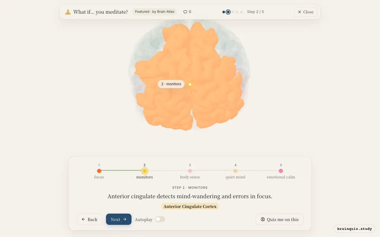
The anterior cingulate cortex (ACC) acts as a conflict monitor, signaling when attention drifts from the intended focus. In meditation, the ACC becomes active when the meditator notices a distraction, prompting a return to the breath. This error-detection process is thought to underlie the 'noticing' aspect of mindfulness.
3. Insula: body sense
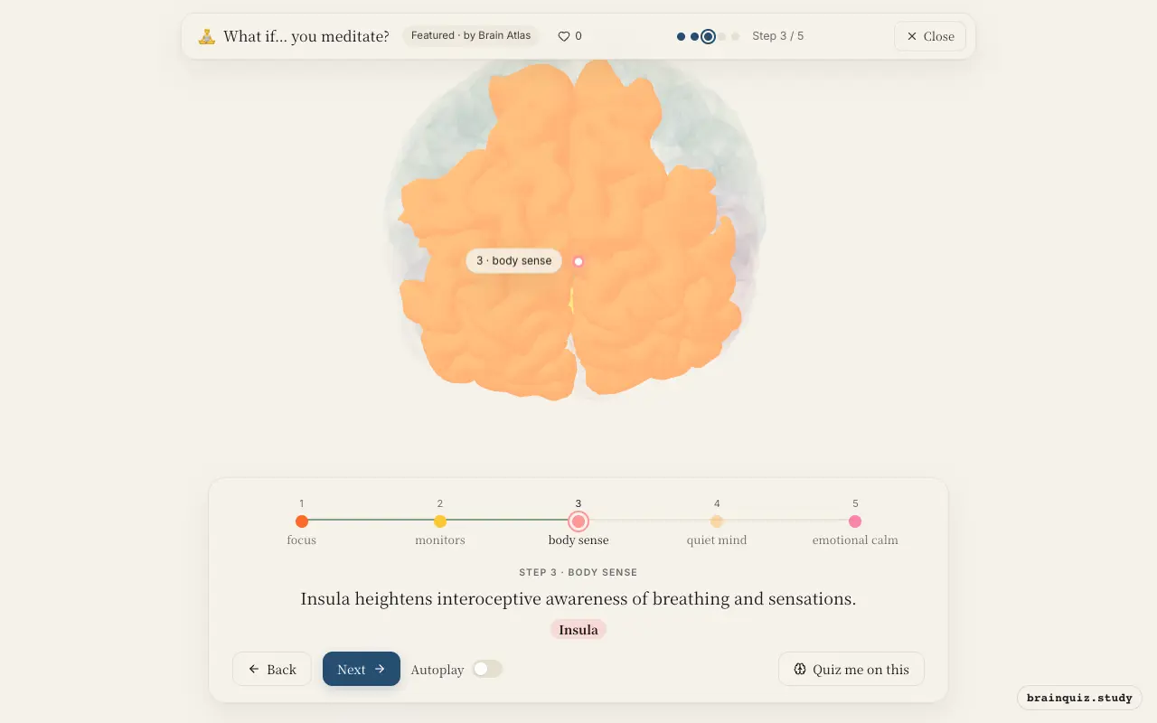
The insula is central to interoception—the perception of internal bodily states. During meditation, attention to the breath or body scan engages the insula, enhancing sensitivity to subtle physiological signals. This improved interoceptive accuracy is linked to better emotional regulation and self-awareness.
4. Posterior Cingulate Cortex: quiet mind
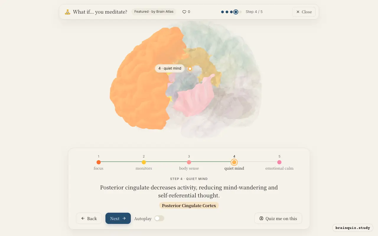
The posterior cingulate cortex is a hub of the default mode network, active during daydreaming and self-referential processing. Focused attention meditation suppresses its activity, which correlates with fewer spontaneous thoughts and a sense of present-centered awareness. Long-term meditators may show less default mode activation even at rest.
5. Amygdala: emotional calm
The amygdala, a key region for detecting threats and generating fear responses, shows reduced activation during meditation, especially in experienced practitioners. This downregulation is associated with lower stress hormones and greater emotional stability. Meditation may strengthen prefrontal-amygdala connections, improving top-down control.
Good to know
- Long-term meditation is associated with increased cortical thickness in prefrontal and insular regions.
- Even brief mindfulness training can reduce amygdala reactivity to emotional stimuli.
- The default mode network, including the posterior cingulate, becomes less active during focused attention meditation.
Meditation vs. Stress Response
Under stress, the amygdala fires and the sympathetic nervous system accelerates heart rate and breathing, preparing for danger. During meditation, the prefrontal cortex and insula become active, downregulating the amygdala and promoting a parasympathetic state of calm and recovery.
Test yourself
Which brain region is primarily responsible for sustaining attention during meditation?
What typically happens to the posterior cingulate cortex during focused attention meditation?
How does meditation affect the amygdala in experienced practitioners?
0 of 3 answered · 0 correct
Simplified: many regions work together at once, and this shows the main players in the order they act. For learning only, not medical advice.