What happens in your brain when you feel disgusted?
Updated
The insula detects the visceral revulsion, the orbitofrontal cortex appraises it as aversive, the anterior cingulate flags conflict, and the putamen drives the grimace and avoidance.
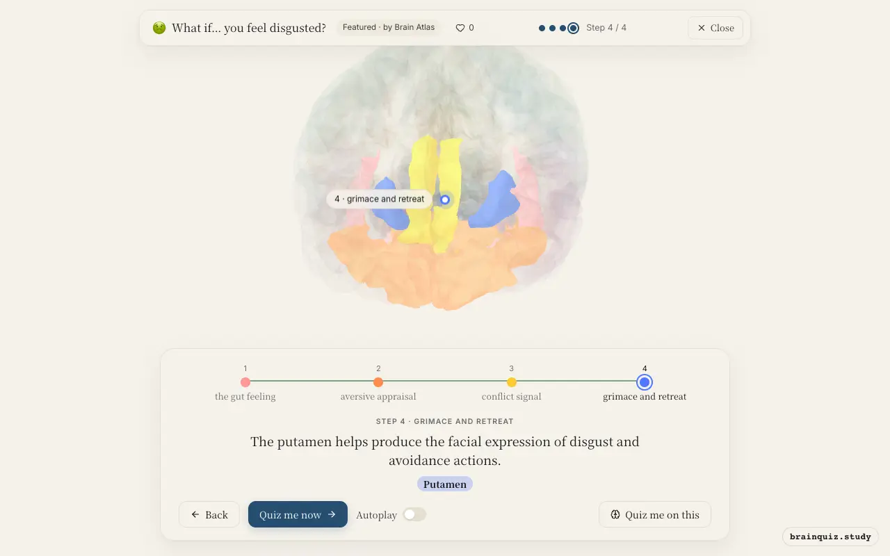
Watch it light up on the 3D brain
Brain regions that light up
| Order | Region | What it does here |
|---|---|---|
| 1 | Insula | The insula fires as the brain registers a visceral sense of revulsion. |
| 2 | Orbitofrontal Cortex | The orbitofrontal cortex evaluates the stimulus as bad or toxic. |
| 3 | Anterior Cingulate Cortex | The anterior cingulate cortex detects the mismatch between expectation and reality. |
| 4 | Putamen | The putamen helps produce the facial expression of disgust and avoidance actions. |
1. Insula: the gut feeling
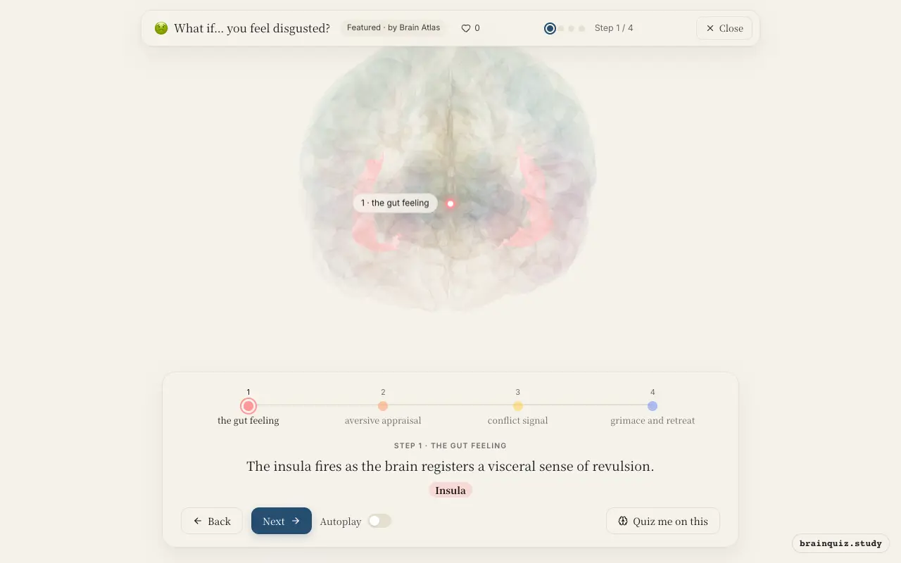
The insula is the hub for interoception—the perception of internal bodily states. During disgust, increased insula activity tracks feelings of nausea and distaste, making it the most consistently activated region in neuroimaging studies of disgust.
2. Orbitofrontal Cortex: aversive appraisal
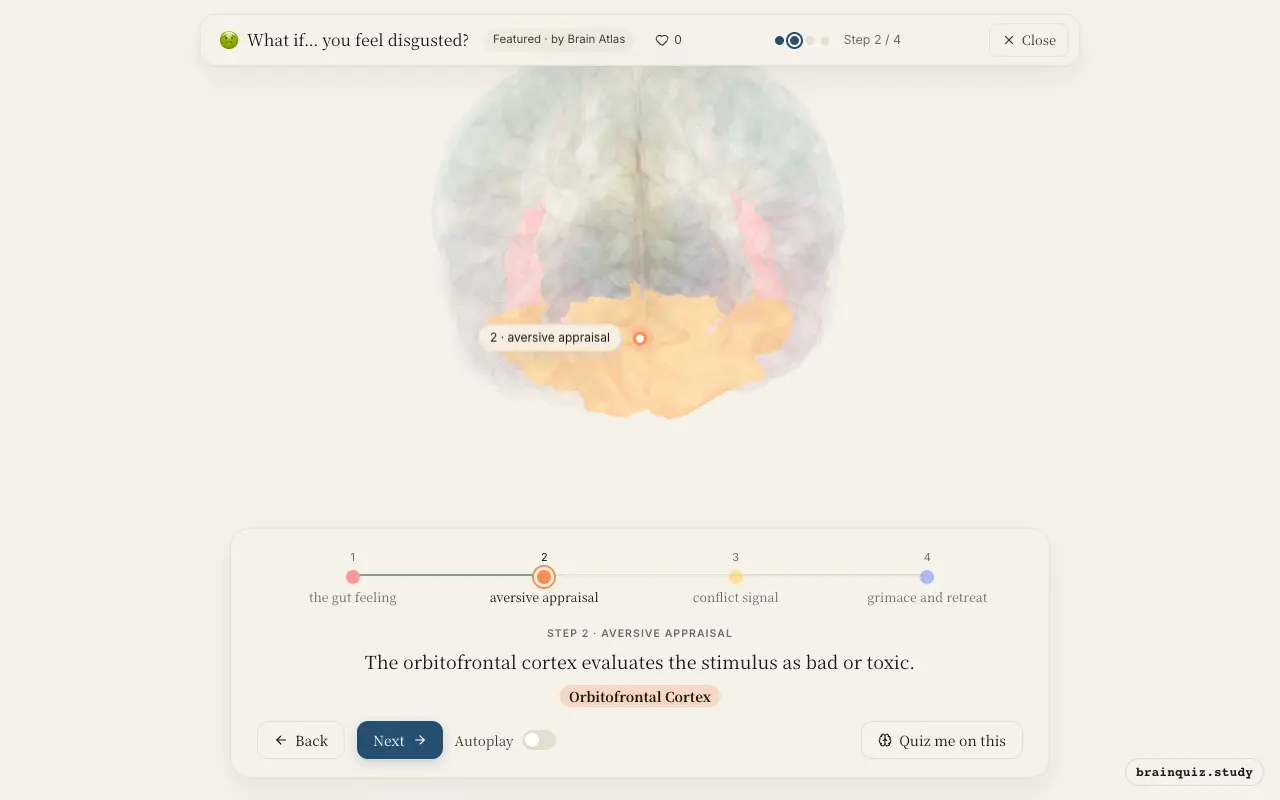
The orbitofrontal cortex computes the emotional and reward value of stimuli. In disgust, it assigns a strongly negative valence, integrating sensory properties with learned aversions to guide avoidance.
3. Anterior Cingulate Cortex: conflict signal
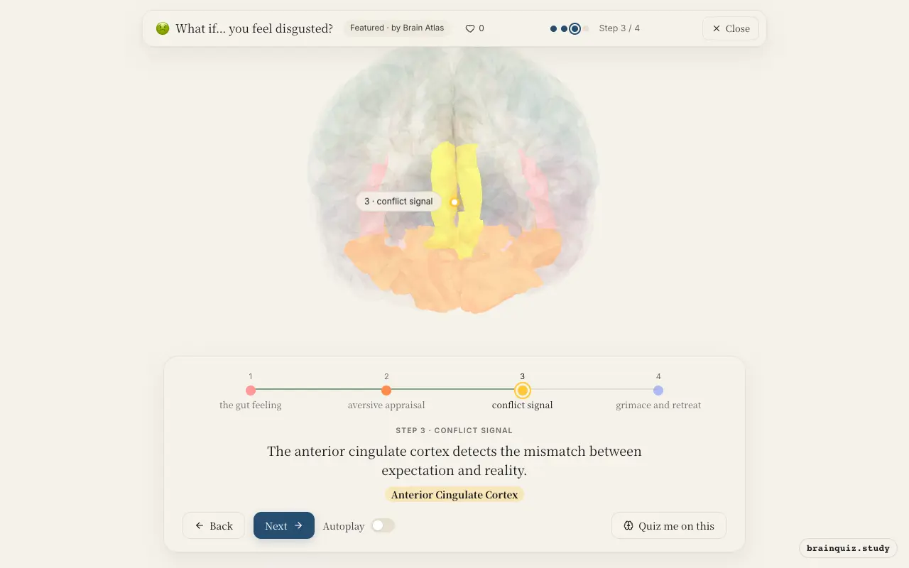
The anterior cingulate cortex monitors conflict and error. Encountering something disgusting engages the ACC, but whether it specifically signals conflict is still debated.
4. Putamen: grimace and retreat
As part of the basal ganglia, the putamen is involved in motor control and habit formation. It coordinates the characteristic wrinkled nose, raised upper lip, and stepping back that accompany disgust.
Good to know
- The insula also responds to viewing disgusted facial expressions in others, supporting empathy.
- Patients with damage to the insula often have difficulty recognizing disgust in others.
- Disgust is thought to have evolved to protect against contamination and disease.
Disgust vs Fear
Fear activates the amygdala as the central threat detector, while disgust relies more on the insula. Both engage the anterior cingulate for salience, but disgust adds strong interoceptive and gustatory processing, and fear adds autonomic fight-or-flight responses.
Test yourself
Which brain region is most consistently activated during the experience of disgust?
What role does the orbitofrontal cortex play in disgust?
Damage to which region impairs recognition of disgust in others?
0 of 3 answered · 0 correct
Simplified: many regions work together at once, and this shows the main players in the order they act.