What happens in your brain when you get dizzy after spinning?
Updated
When you stop spinning, fluid in your inner ear keeps moving, sending signals through the brainstem to the cerebellum and thalamus. The parietal cortex detects a mismatch with vision and body sense, while the insula produces the feeling of dizziness.
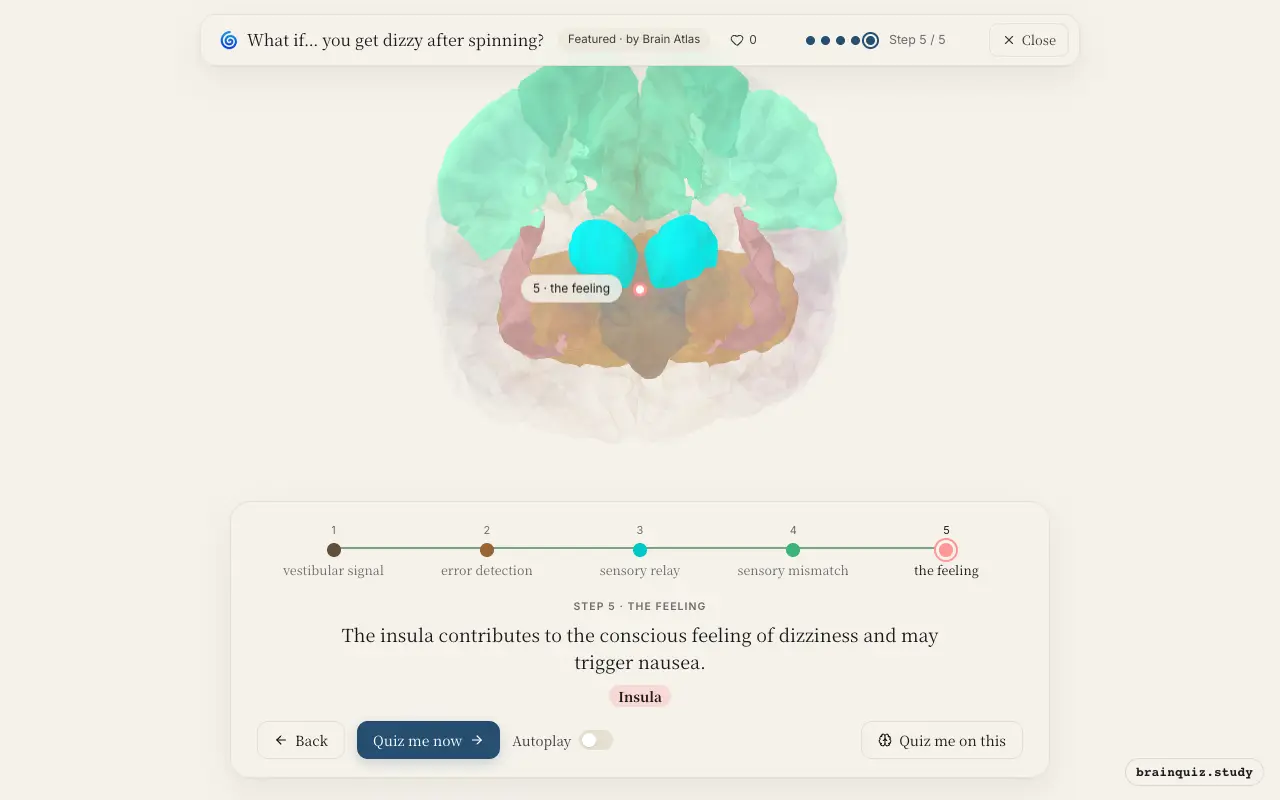
Watch it light up on the 3D brain
Brain regions that light up
| Order | Region | What it does here |
|---|---|---|
| 1 | Brainstem | After spinning stops, inner-ear fluid continues moving, stimulating hair cells that send signals to brainstem vestibular nuclei. |
| 2 | Cerebellum | The cerebellum receives signals and compares them with expected movement, generating corrective commands for eyes and posture. |
| 3 | Thalamus | The thalamus relays vestibular and proprioceptive information to the cerebral cortex, especially the parietal lobe. |
| 4 | Parietal Cortex | The parietal cortex integrates visual, vestibular, and proprioceptive inputs, detecting that they disagree. |
| 5 | Insula | The insula contributes to the conscious feeling of dizziness and may trigger nausea. |
1. Brainstem: vestibular signal
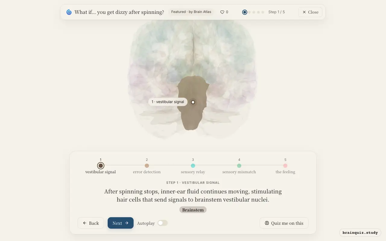
The brainstem houses the vestibular nuclei, which receive input from the semicircular canals. These nuclei integrate information about head rotation and relay it to the cerebellum and thalamus. Because the fluid keeps moving after you stop, the brainstem continues to signal that you are still turning.
2. Cerebellum: error detection
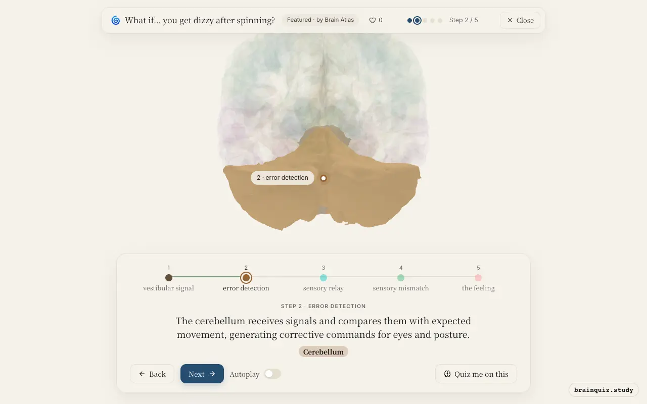
The cerebellum is crucial for motor coordination and balance. It detects the mismatch between the ongoing vestibular signal and the fact that the body has stopped spinning. This triggers adjustments to the vestibulo-ocular reflex and postural muscles, but the conflict persists as long as the fluid moves.
3. Thalamus: sensory relay
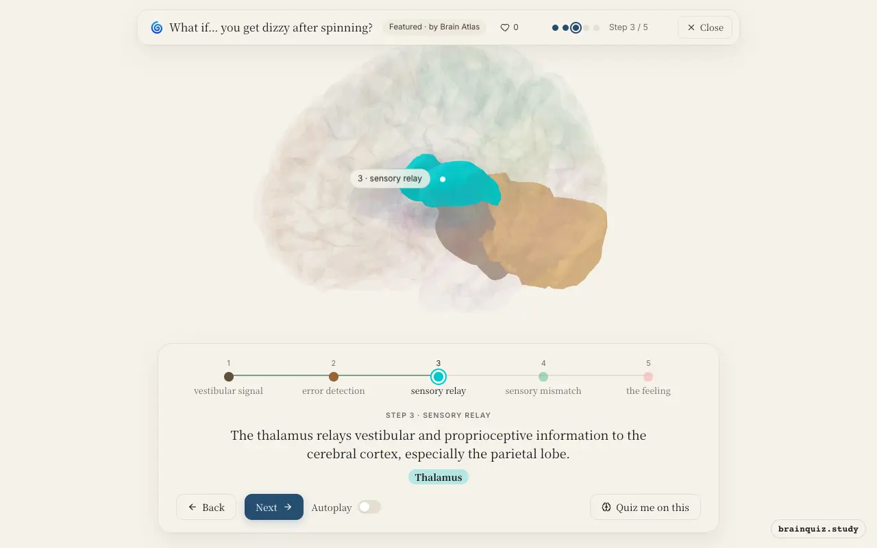
As the brain's main sensory relay station, the thalamus routes signals from the brainstem to the cortex. It ensures that the parietal cortex receives the vestibular input alongside visual and somatosensory information. This integration is essential for perceiving one's body position in space.
4. Parietal Cortex: sensory mismatch
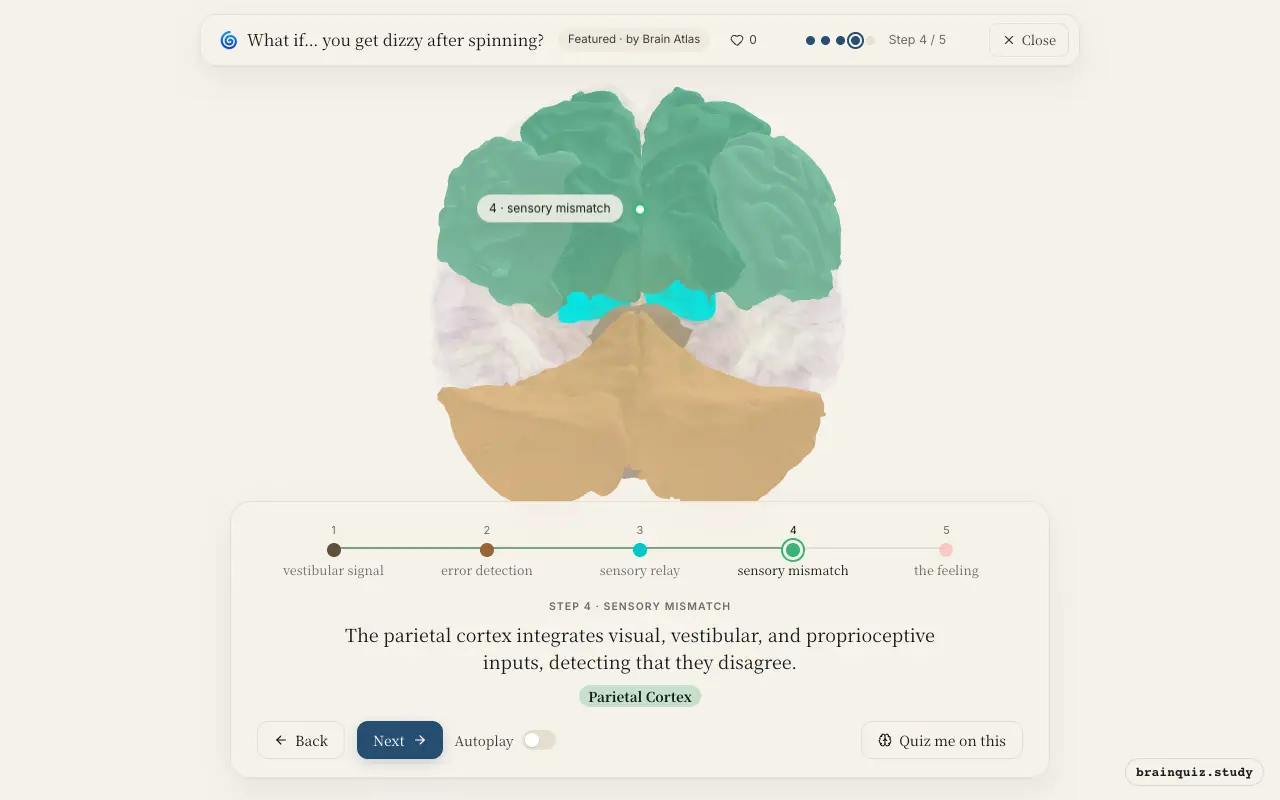
The parietal cortex, particularly the right posterior parietal area, constructs a sense of body orientation. It compares the vestibular signal of continued rotation with visual input showing a stationary world and proprioceptive input indicating no movement. This conflict is thought to produce the sensation of dizziness.
5. Insula: the feeling
The insula processes interoceptive signals from the body, including those from the vestibular system. It is thought to generate the subjective experience of dizziness and related autonomic responses like nausea. Activity in the anterior insula is often associated with such unpleasant bodily sensations.
Good to know
- The semicircular canals detect angular acceleration, and their fluid (endolymph) has inertia, so it keeps moving after rotation stops.
- The vestibulo-ocular reflex, coordinated by the cerebellum, causes nystagmus (involuntary eye movements) during and after spinning.
- Dizziness after spinning is a form of sensory conflict, similar to motion sickness, where the brain receives contradictory motion signals.
Post-spin dizziness vs. BPPV vertigo
Post-spin dizziness is triggered by normal fluid inertia after voluntary rotation, while BPPV involves displaced otoconia causing false signals with head movements. Brain activity is similar in vestibular and cerebellar regions, but BPPV episodes are typically shorter and position-dependent, whereas post-spin dizziness resolves as the fluid settles.
Test yourself
Which brain structure first receives signals from the inner ear after spinning stops?
What role does the cerebellum play in post-spin dizziness?
Which region is thought to produce the conscious sensation of dizziness?
0 of 3 answered · 0 correct
Simplified: many regions work together at once, and this shows the main players in the order they act.