What happens in your brain when you have depression?
Updated
Depression involves an overactive amygdala for negative emotions, a weakened prefrontal cortex that fails to regulate it, a blunted nucleus accumbens reducing pleasure, and a reduced-volume hippocampus impairing memory.
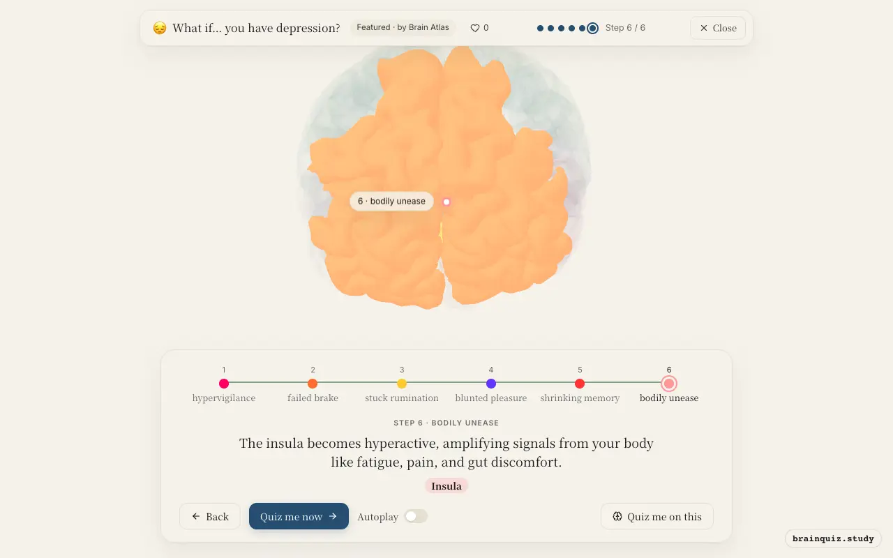
Watch it light up on the 3D brain
Brain regions that light up
| Order | Region | What it does here |
|---|---|---|
| 1 | Amygdala | The amygdala fires more strongly to negative faces and memories, coloring your world with threat. |
| 2 | Prefrontal Cortex | The prefrontal cortex, especially dorsolateral parts, is underactive, so it can't dampen the amygdala's alarm. |
| 3 | Anterior Cingulate Cortex | The anterior cingulate cortex gets caught in loops of self-focus and error monitoring, feeding rumination. |
| 4 | Nucleus Accumbens | The nucleus accumbens responds weakly to rewards, so everyday joys lose their shine. |
| 5 | Hippocampus | Chronic stress may shrink the hippocampus, impairing memory and reinforcing depressive thinking. |
| 6 | Insula | The insula becomes hyperactive, amplifying signals from your body like fatigue, pain, and gut discomfort. |
1. Amygdala: hypervigilance
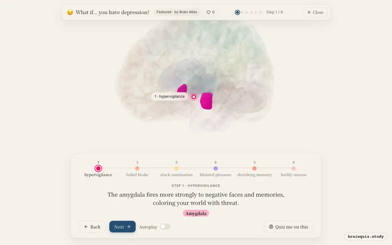
In depression, the amygdala shows exaggerated responses to sad or fearful stimuli and even to neutral cues. This overactivity biases perception toward negative interpretations and sustains a low mood. It also drives the stress response, creating a vicious cycle. This reactivity is thought to reflect reduced inhibitory control from the prefrontal cortex.
2. Prefrontal Cortex: failed brake
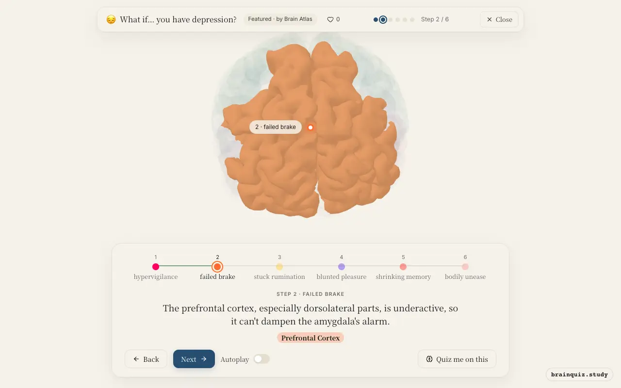
Normally, the prefrontal cortex exerts top-down control over emotional responses, but in depression its activity is reduced, particularly in the dorsolateral and ventromedial regions. This weakens the ability to reappraise negative thoughts and regulate mood, leaving the amygdala unchecked. Consequently, negative emotions persist and cognitive flexibility declines.
3. Anterior Cingulate Cortex: stuck rumination
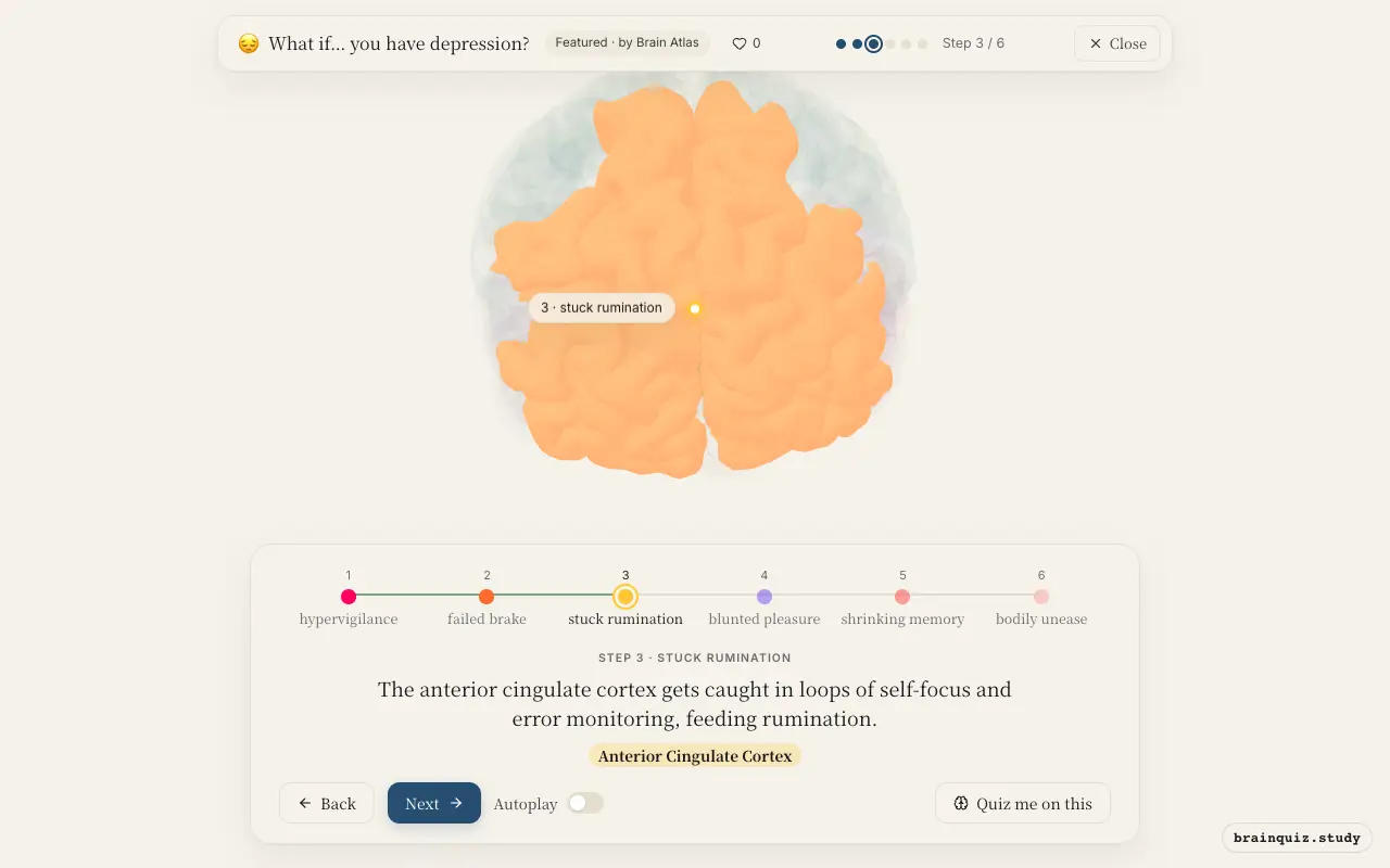
The anterior cingulate cortex, involved in conflict monitoring and error detection, shows abnormal activity in depression, often with increased rostral ACC during negative self-referential processing. This may drive rumination—repetitive negative thinking—and contribute to the feeling that something is wrong. It also connects emotional and cognitive networks, amplifying distress.
4. Nucleus Accumbens: blunted pleasure
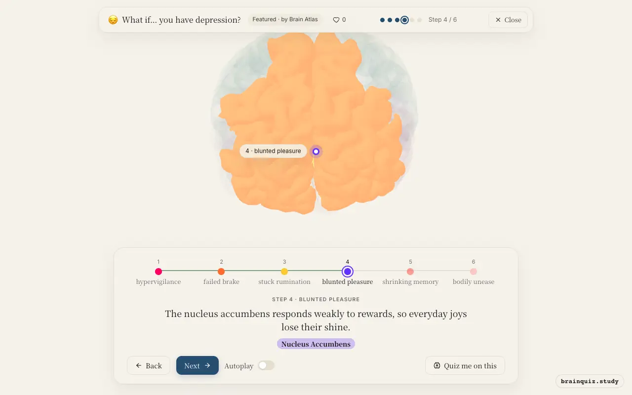
The nucleus accumbens is a hub for reward and motivation, but in depression its response to positive stimuli is blunted, likely due to reduced dopamine signaling from the ventral tegmental area. This underlies anhedonia—the inability to feel pleasure—and saps the drive to pursue goals. Even previously enjoyable activities no longer feel rewarding.
5. Hippocampus: shrinking memory
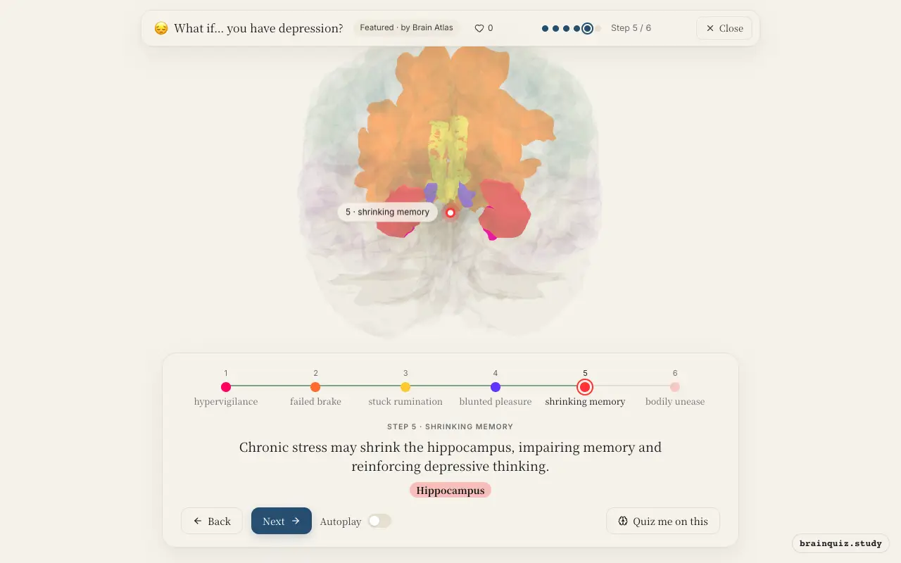
The hippocampus is sensitive to stress hormones like cortisol, and in depression it often shows reduced volume, possibly from prolonged exposure to elevated glucocorticoids. This impairs memory formation and contextual processing, making it harder to recall positive experiences. A smaller hippocampus may also contribute to the cognitive symptoms of depression, such as poor concentration.
6. Insula: bodily unease
The insula integrates interoceptive signals—sensations from inside the body—and in depression its activity is heightened, leading to a heightened awareness of physical symptoms like fatigue, aches, and changes in appetite. This can create a feedback loop where bodily discomfort worsens mood, and negative emotions intensify bodily sensations. The insula also plays a role in self-awareness and empathy, which may be altered.
Good to know
- Depression is associated with elevated cortisol levels, which may contribute to hippocampal volume loss.
- Successful treatment with antidepressants or psychotherapy is thought to increase neuroplasticity in the prefrontal cortex and hippocampus.
- Genetic and environmental factors interact to influence the development of depression.
Depression vs. Sadness
Normal sadness involves transient amygdala and prefrontal changes that return to baseline, while depression shows persistent overactivity in the amygdala, underactivity in the prefrontal cortex, and blunted reward response in the nucleus accumbens. The hippocampus may also shrink in chronic depression but not in brief sadness.
Test yourself
Which brain region is typically overactive in depression, contributing to negative emotional bias?
What is the likely consequence of reduced nucleus accumbens activity in depression?
How does the hippocampus change in many people with depression?
0 of 3 answered · 0 correct
Simplified: many regions work together at once, and this shows the main players in the order they act. For learning only, not medical advice.