What happens in your brain when you lift weights?
Updated
Your prefrontal cortex plans the lift, premotor cortex sequences it, basal ganglia initiate the movement, motor cortex commands muscles, cerebellum fine-tunes, and somatosensory cortex processes feedback.
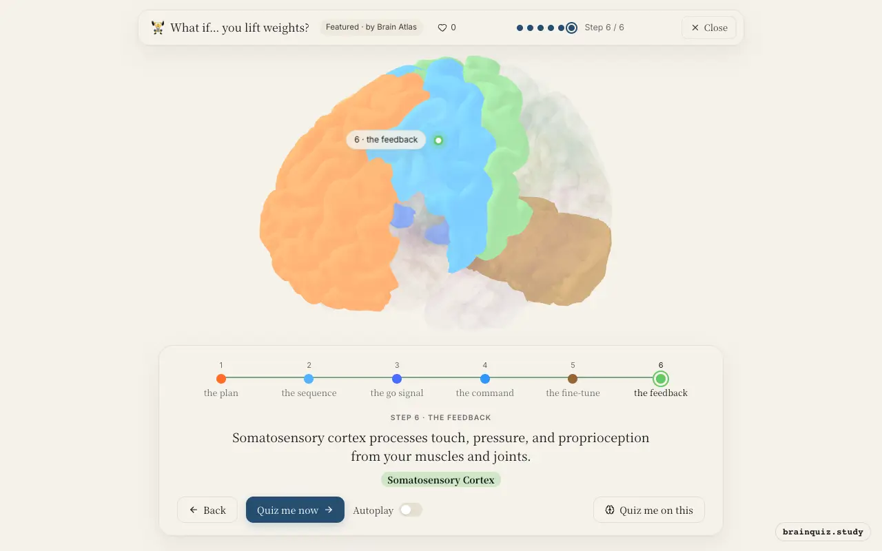
Watch it light up on the 3D brain
Brain regions that light up
| Order | Region | What it does here |
|---|---|---|
| 1 | Prefrontal Cortex | You decide to lift and set the goal; prefrontal cortex focuses attention on the task. |
| 2 | Premotor Cortex | Premotor cortex assembles the sequence of muscle activations needed for the lift. |
| 3 | Basal Ganglia | Basal ganglia help select and initiate the movement, releasing inhibition on the thalamus. |
| 4 | Motor Cortex | Motor cortex sends signals down the corticospinal tract to activate muscles. |
| 5 | Cerebellum | Cerebellum compares intended movement with actual sensory feedback and corrects errors. |
| 6 | Somatosensory Cortex | Somatosensory cortex processes touch, pressure, and proprioception from your muscles and joints. |
1. Prefrontal Cortex: the plan
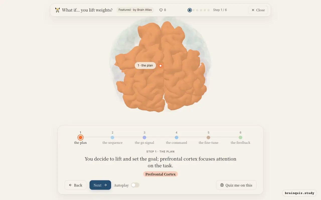
The prefrontal cortex, especially dorsolateral parts, is active when you intentionally initiate a motor act. It holds the goal in working memory, suppresses distractions, and signals your intention to move. Studies suggest it sends top-down input to premotor and motor areas.
2. Premotor Cortex: the sequence
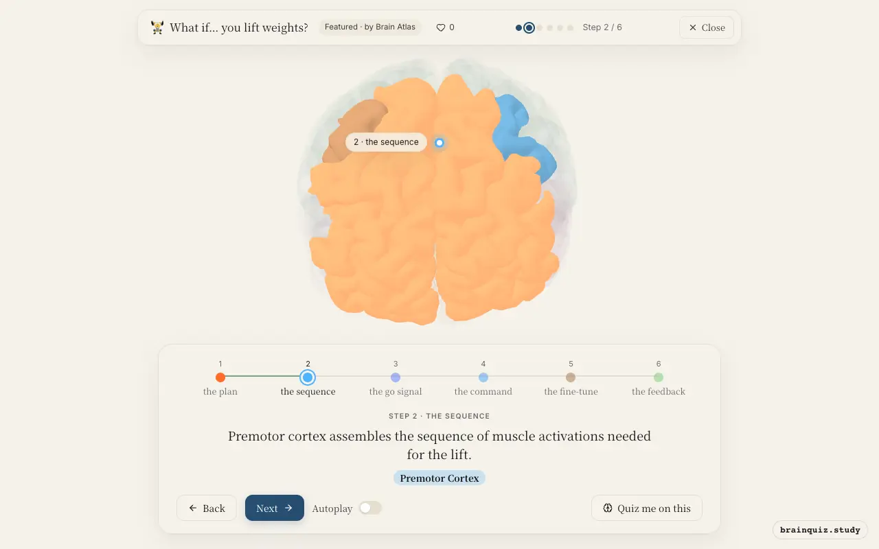
The premotor cortex uses sensory information and past experience to plan the movement trajectory. It prepares the motor program before execution, integrating spatial and body position cues. This planning is essential for coordinating multiple joints and muscles.
3. Basal Ganglia: the go signal
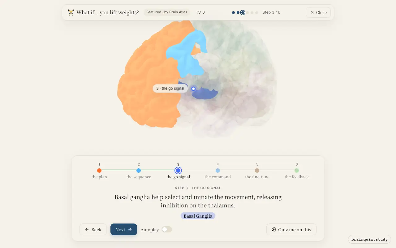
The basal ganglia act as a gate for voluntary movement. The direct pathway disinhibits the thalamus, allowing motor commands to proceed. This selection process ensures the correct movement is chosen and scaled appropriately.
4. Motor Cortex: the command
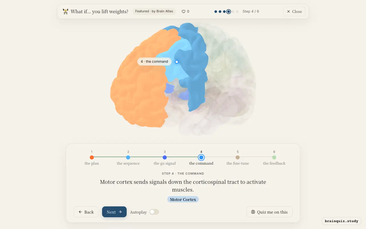
The primary motor cortex contains a somatotopic map of the body. Neurons here fire to produce force and direction of movement. The signal travels via upper motor neurons to lower motor neurons in the spinal cord, causing muscle contraction.
5. Cerebellum: the fine-tune
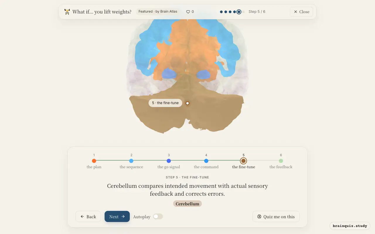
The cerebellum receives a copy of the motor command (efference copy) and sensory input from muscles and joints. It computes the difference between intended and actual movement, then sends corrective signals to the motor cortex and brainstem. This ensures smooth, coordinated lifting.
6. Somatosensory Cortex: the feedback
As you lift, receptors in muscles, tendons, and joints send signals via the spinal cord and thalamus to the somatosensory cortex. This region creates awareness of limb position and load, allowing you to adjust force if needed. It is critical for maintaining proper form.
Good to know
- The corticospinal tract decussates at the medullary pyramids, meaning the left motor cortex controls the right side of the body.
- Motor learning from repeated lifting leads to long-term potentiation in the motor cortex and cerebellum, making movements more efficient.
- The basal ganglia's direct pathway promotes movement by releasing the thalamus from tonic inhibition.
Lifting vs. Imagining Lifting
Imagining a lift activates prefrontal and premotor cortex and weakly activates primary motor cortex, so no movement occurs. Actual lifting strongly drives motor cortex, cerebellum, and basal ganglia output, along with ongoing somatosensory feedback to correct form.
Test yourself
Which region is primarily responsible for planning the sequence of muscle activations before you lift?
The cerebellum's role during weightlifting is best described as:
What is the function of the basal ganglia in voluntary movement like lifting?
0 of 3 answered · 0 correct
Simplified: many regions work together at once, and this shows the main players in the order they act.