What happens in your brain when you read a map?
Updated
The visual cortex decodes map symbols, the angular gyrus processes spatial relationships, the parahippocampal gyrus recognizes the map as a scene, the hippocampus retrieves spatial memories, and the prefrontal cortex plans the route.
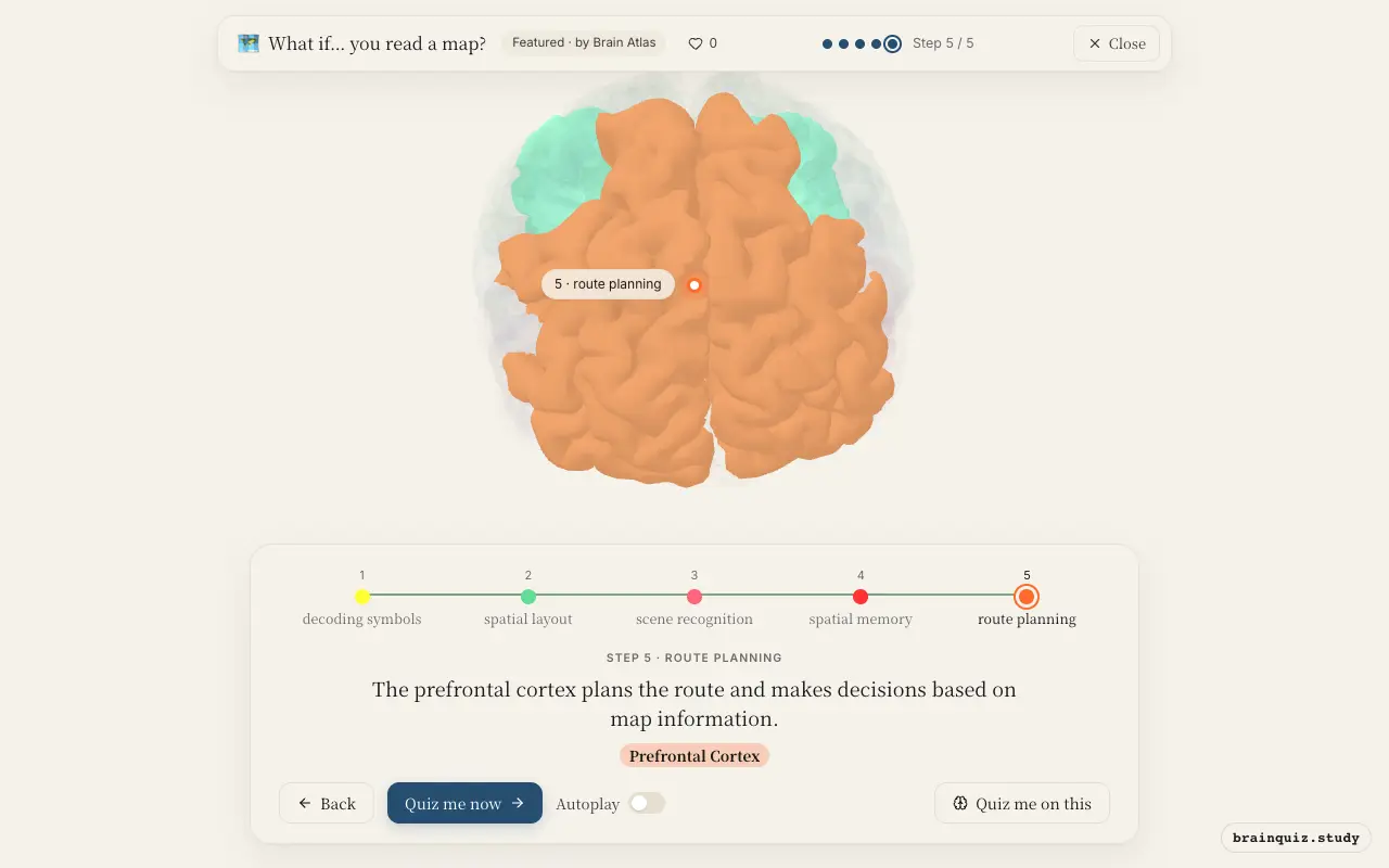
Watch it light up on the 3D brain
Brain regions that light up
| Order | Region | What it does here |
|---|---|---|
| 1 | Visual Cortex | The primary visual cortex processes the map's lines, colors, and symbols from the retina. |
| 2 | Angular Gyrus | The angular gyrus integrates visual input to understand spatial relationships and map orientation. |
| 3 | Parahippocampal Gyrus | The parahippocampal gyrus identifies the map as a spatial scene, not just a flat image. |
| 4 | Hippocampus | The hippocampus compares the map with stored spatial memories to recognize familiar places. |
| 5 | Prefrontal Cortex | The prefrontal cortex plans the route and makes decisions based on map information. |
1. Visual Cortex: decoding symbols
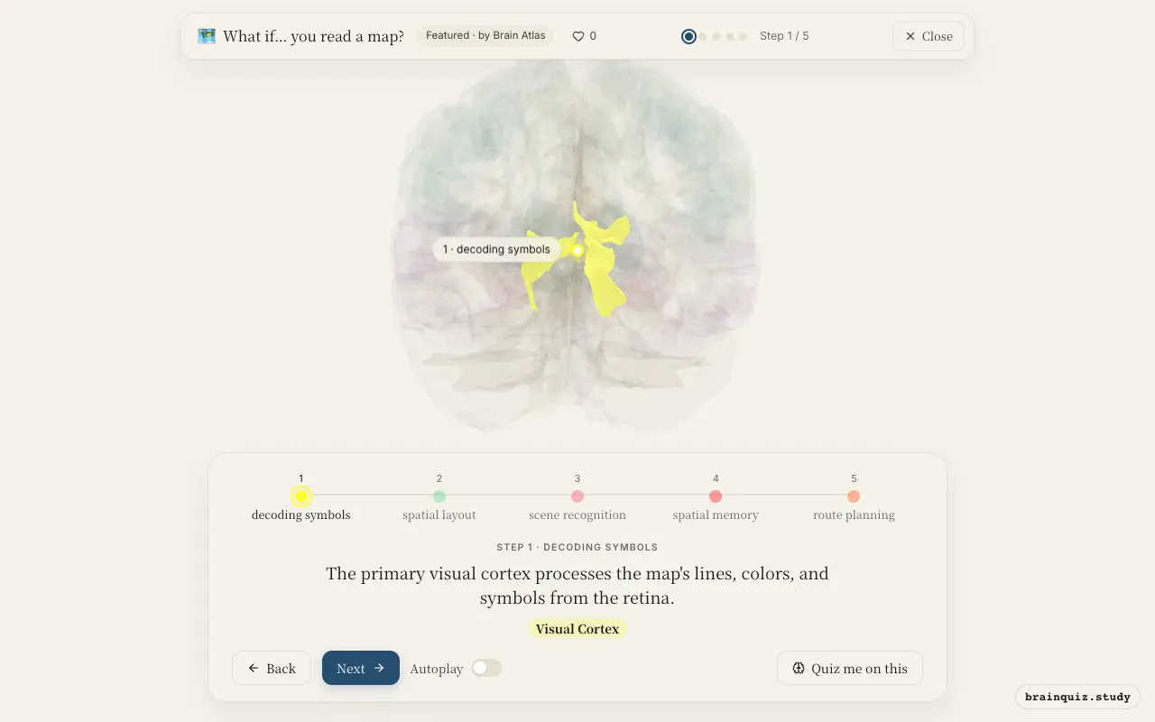
The visual cortex in the occipital lobe receives raw visual input and extracts basic features like edges, orientations, and contrast. This initial analysis extracts features that higher-order areas use to identify map elements such as roads, landmarks, and text labels. Without this step, the map would be an unrecognizable pattern.
2. Angular Gyrus: spatial layout
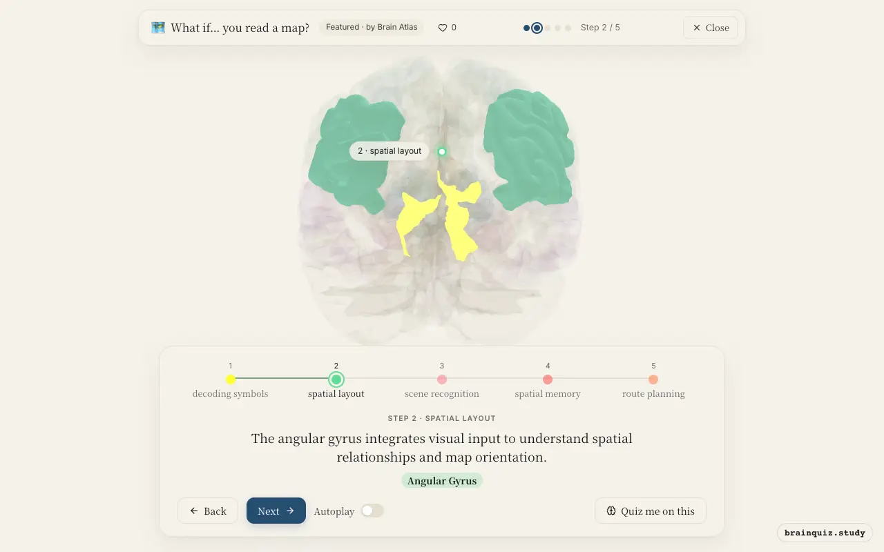
Located in the posterior parietal cortex, the angular gyrus is crucial for spatial cognition and mental rotation. It helps you interpret the relative positions of map features and align the map with your real-world orientation, a process known as perspective-taking. Damage here can cause difficulties in map reading and navigation.
3. Parahippocampal Gyrus: scene recognition
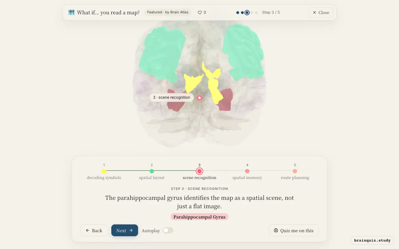
The parahippocampal place area (PPA) within the parahippocampal gyrus responds selectively to scenes and spatial layouts. It categorizes the map as a representation of a navigable environment, allowing subsequent brain regions to retrieve or form spatial memories. This region helps distinguish maps from other visual objects like faces or tools.
4. Hippocampus: spatial memory
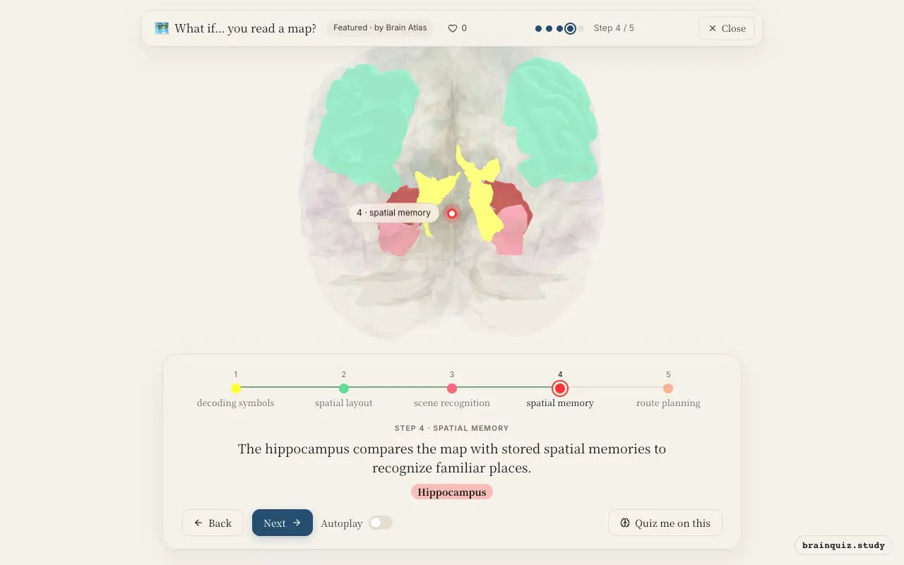
The hippocampus is essential for spatial navigation and memory consolidation. It contains place cells that fire when you are in a specific location, and pre-existing cognitive maps stored here can be matched to the map's layout. If you have explored the area before, the hippocampus retrieves that knowledge to help you navigate.
5. Prefrontal Cortex: route planning
The prefrontal cortex, especially the dorsolateral part, integrates spatial information with goals and constraints to devise a navigation strategy. It weighs alternatives, such as the fastest versus the most scenic path, and holds the plan in working memory while you execute it. This executive control is needed when the route is unfamiliar or complex.
Good to know
- The parahippocampal place area responds selectively to scenes and spatial layouts, not to isolated objects.
- London taxi drivers have enlarged posterior hippocampi, a finding linked to their extensive spatial navigation experience.
- Mental rotation, a core skill in map reading, consistently activates the parietal cortex, including the angular gyrus.
Map reading vs. GPS navigation
Reading a map requires active spatial processing in the parietal cortex and hippocampus, engaging mental rotation and memory retrieval. In contrast, following GPS instructions may rely more on working memory and language areas, as auditory commands replace visual-spatial analysis. Thus, map readers may build stronger cognitive maps than GPS users.
Test yourself
Which brain region is primarily responsible for recognizing a map as a spatial scene?
What role does the hippocampus play in map reading?
Which region is most active when you mentally rotate a map to align it with your orientation?
0 of 3 answered · 0 correct
Simplified: many regions work together at once, and this shows the main players in the order they act.