What happens in your brain when you see someone in pain?
Updated
Your visual cortex processes the painful sight; the insula maps your own body state, the anterior cingulate cortex flags distress, and the prefrontal cortex interprets meaning and perspective.
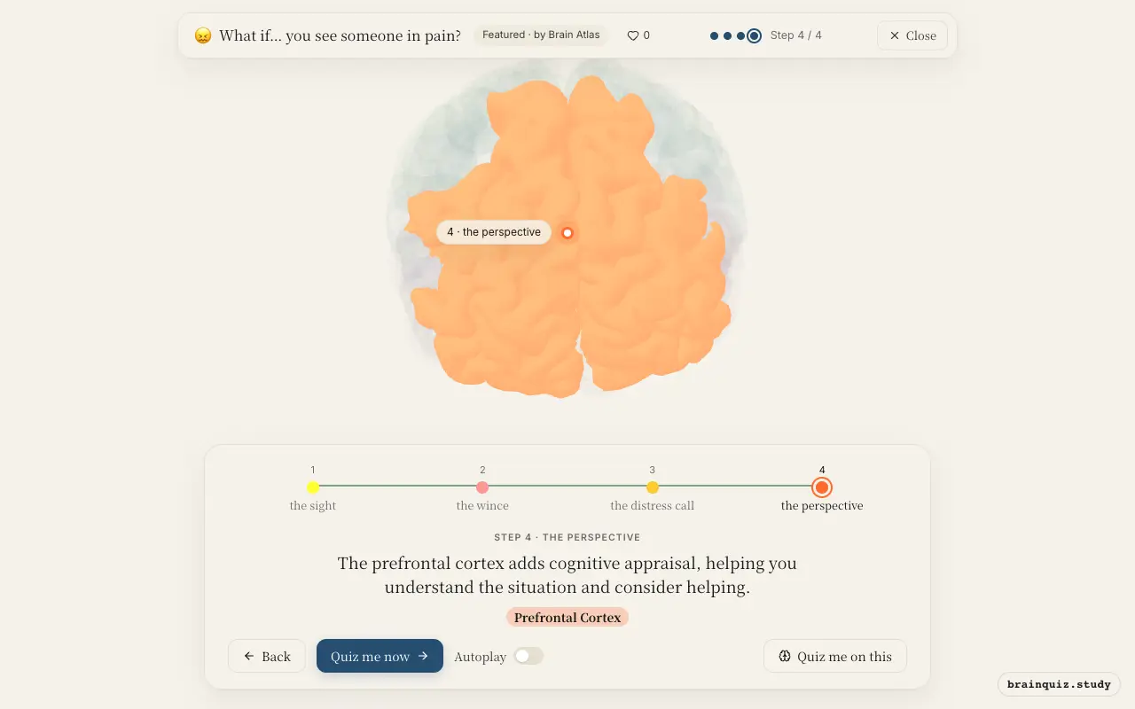
Watch it light up on the 3D brain
Brain regions that light up
| Order | Region | What it does here |
|---|---|---|
| 1 | Visual Cortex | Your primary visual cortex breaks down the image of the other person's pained expression into basic visual features. |
| 2 | Insula | The insula maps your own body signals, creating a shared representation of the other person's pain. |
| 3 | Anterior Cingulate Cortex | Your anterior cingulate cortex registers the negative emotional salience, making the other's pain feel urgent. |
| 4 | Prefrontal Cortex | The prefrontal cortex adds cognitive appraisal, helping you understand the situation and consider helping. |
1. Visual Cortex: the sight
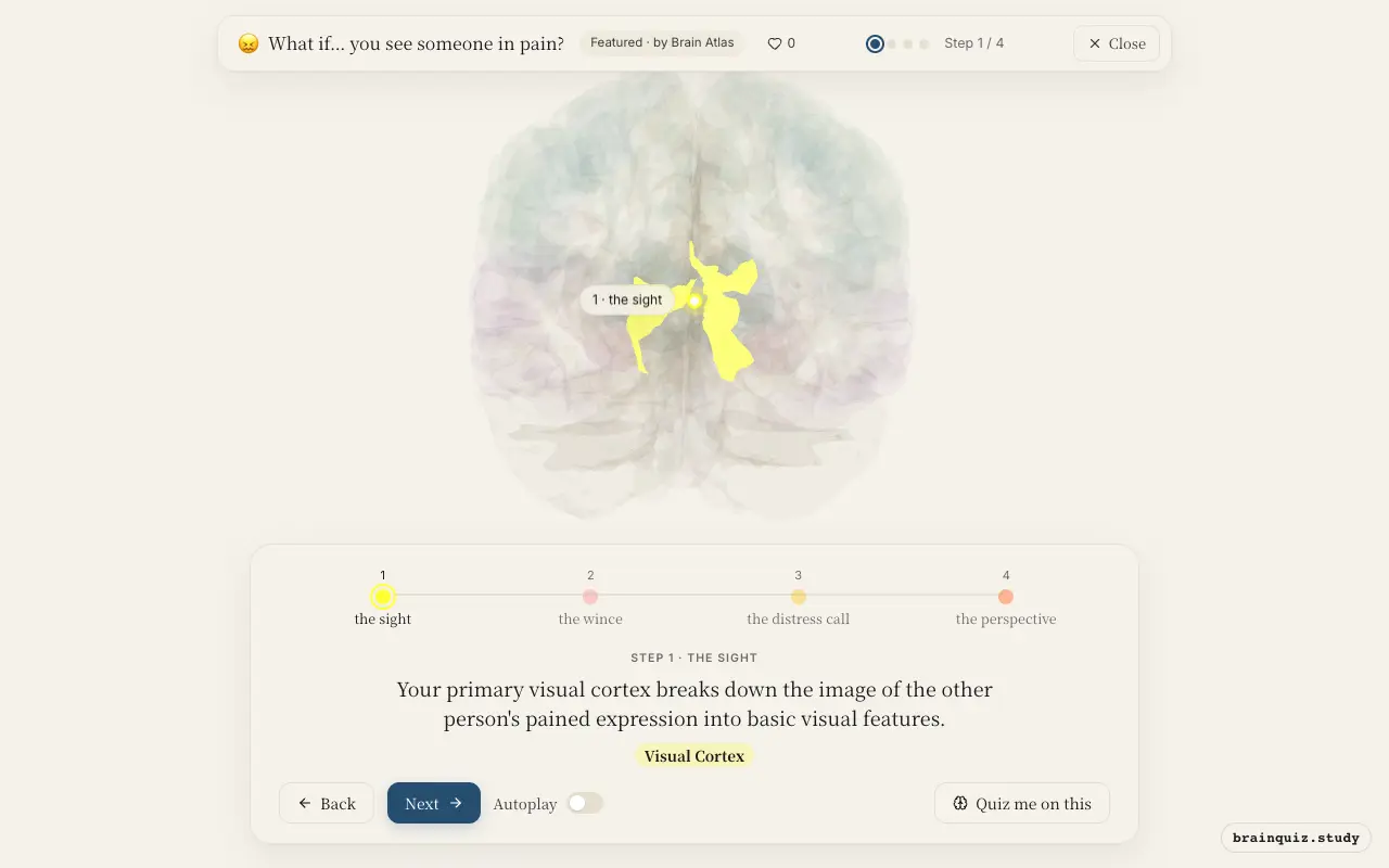
This region is the first cortical stop for visual information, processing edges, motion, and orientation. It constructs the initial representation of the painful scene, which is then sent forward to higher areas for interpretation and emotional tagging.
2. Insula: the wince
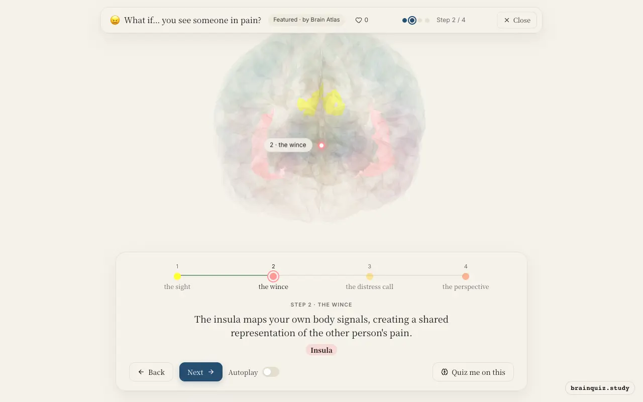
The insula is critical for interoception and empathy. It simulates the other's pain by referencing your own bodily states, producing a visceral, felt sense of their distress. This region is consistently active when witnessing pain in others.
3. Anterior Cingulate Cortex: the distress call
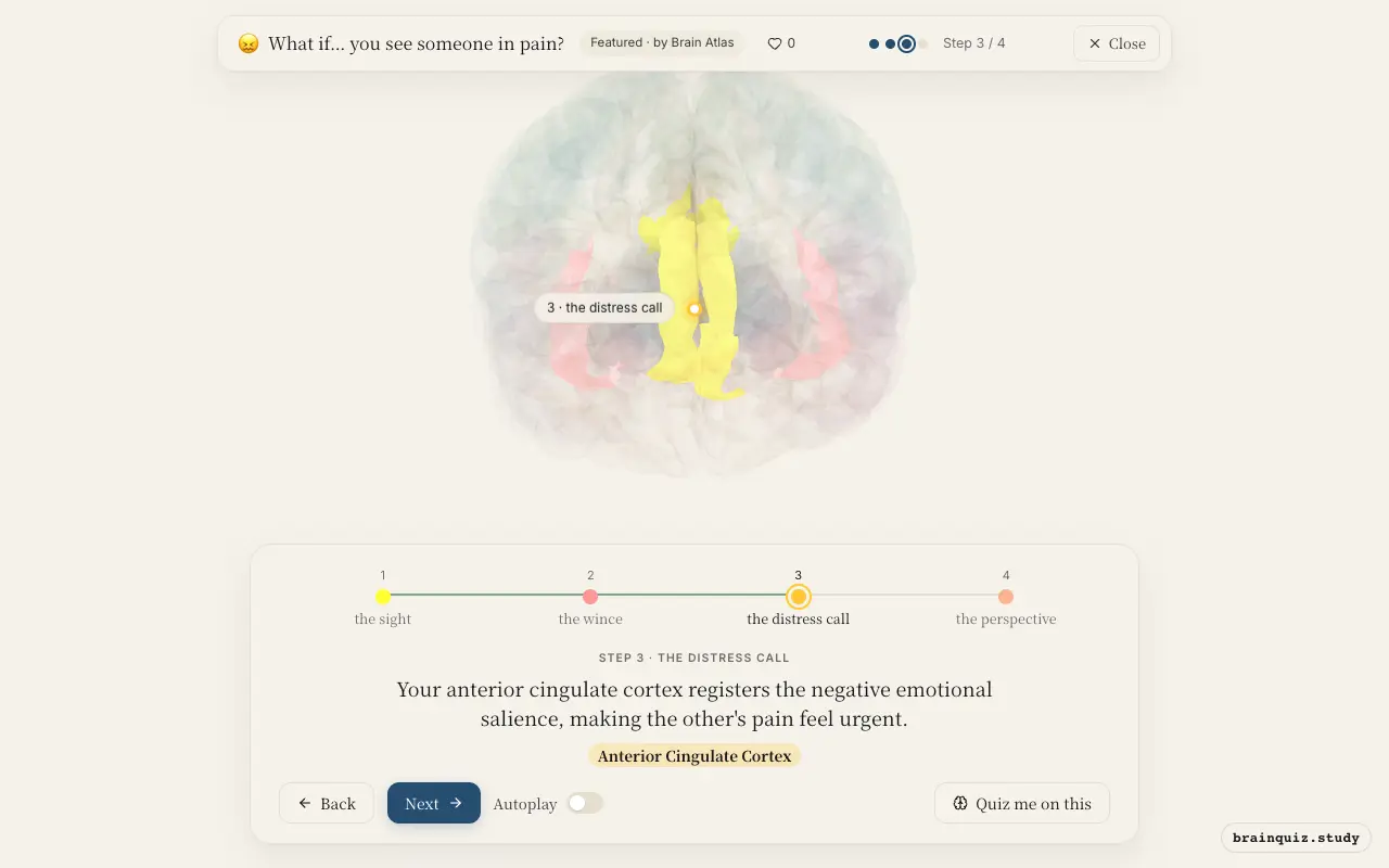
The anterior cingulate is involved in error detection and the affective component of pain. Here, it flags the aversive significance of the other's injury, motivating concern and a potential response.
4. Prefrontal Cortex: the perspective
The prefrontal cortex supports perspective-taking and emotion regulation. It allows you to interpret the painful event in context, weigh appropriate reactions, and plan a prosocial response rather than simply mirroring distress.
Good to know
- The insula and anterior cingulate are often co-activated when witnessing others in pain, forming the core of the 'pain empathy network.'
- People with higher empathy scores show stronger activation in these regions.
- The mirror neuron system, including premotor and parietal areas, may also contribute, but its role is debated.
Seeing pain vs feeling pain yourself
When you feel pain yourself, the somatosensory cortex and thalamus are strongly engaged for sensory detail. When you see pain, activity shifts to the insula and anterior cingulate, reflecting emotional and interoceptive simulation rather than raw sensation.
Test yourself
Which brain region first processes the visual information of someone in pain?
What is the primary role of the insula in empathy for pain?
Which two regions are most consistently activated when witnessing someone else's pain?
0 of 3 answered · 0 correct
Simplified: many regions work together at once, and this shows the main players in the order they act.