What happens in your brain when you see something move?
Updated
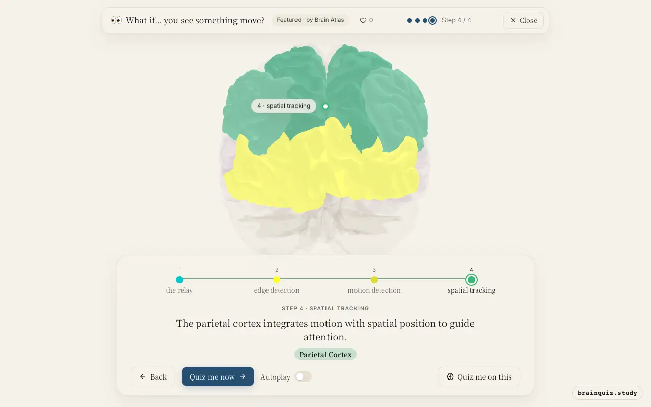
Motion perception begins in the thalamus relaying visual input to the primary visual cortex, which detects edges. The occipital cortex's motion area MT computes direction and speed, and the parietal cortex integrates this with spatial attention.
Watch it light up on the 3D brain
Brain regions that light up
| Order | Region | What it does here |
|---|---|---|
| 1 | Thalamus | The thalamus relays visual signals from the retina to the primary visual cortex. |
| 2 | Visual Cortex | The primary visual cortex (V1) detects oriented edges and basic features. |
| 3 | Occipital Cortex | Area MT in the occipital cortex computes direction and speed of motion. |
| 4 | Parietal Cortex | The parietal cortex integrates motion with spatial position to guide attention. |
1. Thalamus: the relay

The lateral geniculate nucleus of the thalamus receives input from retinal ganglion cells and sends it to V1. This relay preserves retinotopic organization and begins segregating motion and color channels.
2. Visual Cortex: edge detection
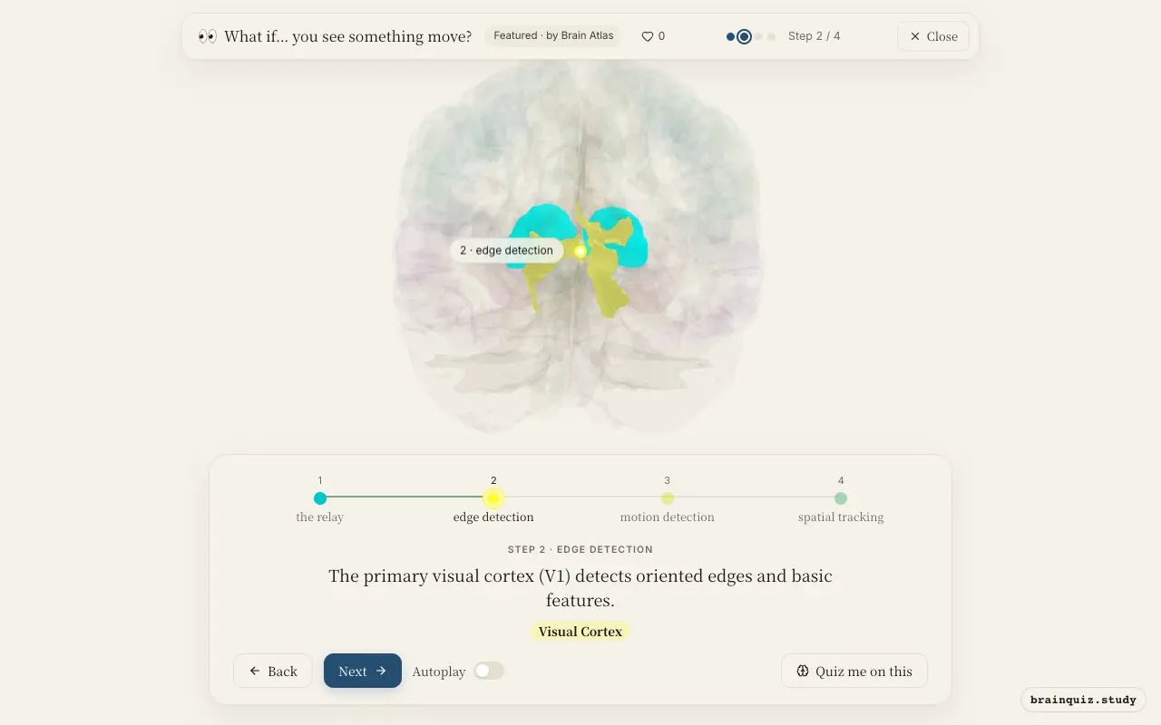
V1 neurons respond to edges, orientation, and spatial frequency. They provide input to higher visual areas that specialize in motion, such as area MT in the occipital cortex.
3. Occipital Cortex: motion detection
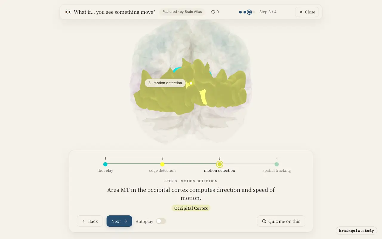
Area MT, located in the occipital cortex, contains direction-selective neurons that respond to specific velocities. It integrates local motion signals from V1 to represent global motion of objects and patterns, enabling perception of movement. Damage to this area can cause akinetopsia, an inability to perceive motion.
4. Parietal Cortex: spatial tracking
The posterior parietal cortex combines motion direction with spatial coordinates to guide attention and eye movements toward the moving object. It is a key node in the dorsal 'where/how' pathway, helping us locate and interact with moving stimuli. This integration supports smooth pursuit eye movements when tracking.
Good to know
- Motion-sensitive area MT (V5) is located in the extrastriate occipital cortex and is highly conserved across primates.
- The dorsal visual stream extends from V1 through MT to the parietal cortex and is specialized for spatial vision and motion.
- Patients with bilateral damage to MT experience akinetopsia, the inability to perceive motion.
Static vs Moving Scene
Viewing a static scene activates primary visual cortex and ventral areas for object recognition, but motion areas like MT stay relatively quiet. When something moves, MT and parietal cortex show strong responses, directing attention and eye movements. The dynamic input transforms a stable picture into a tracking task.
Test yourself
Which region first receives visual input from the retina before it reaches the cortex?
Which area is specialized for detecting the direction and speed of visual motion?
A patient with damage to area MT would most likely have difficulty:
0 of 3 answered · 0 correct
Simplified: many regions work together at once, and this shows the main players in the order they act.