What happens in your brain when you serve in tennis?
Updated
The prefrontal cortex plans the serve, the premotor cortex sequences the toss and swing, the motor cortex executes the movement, the cerebellum coordinates timing, and the basal ganglia ensures smooth execution.
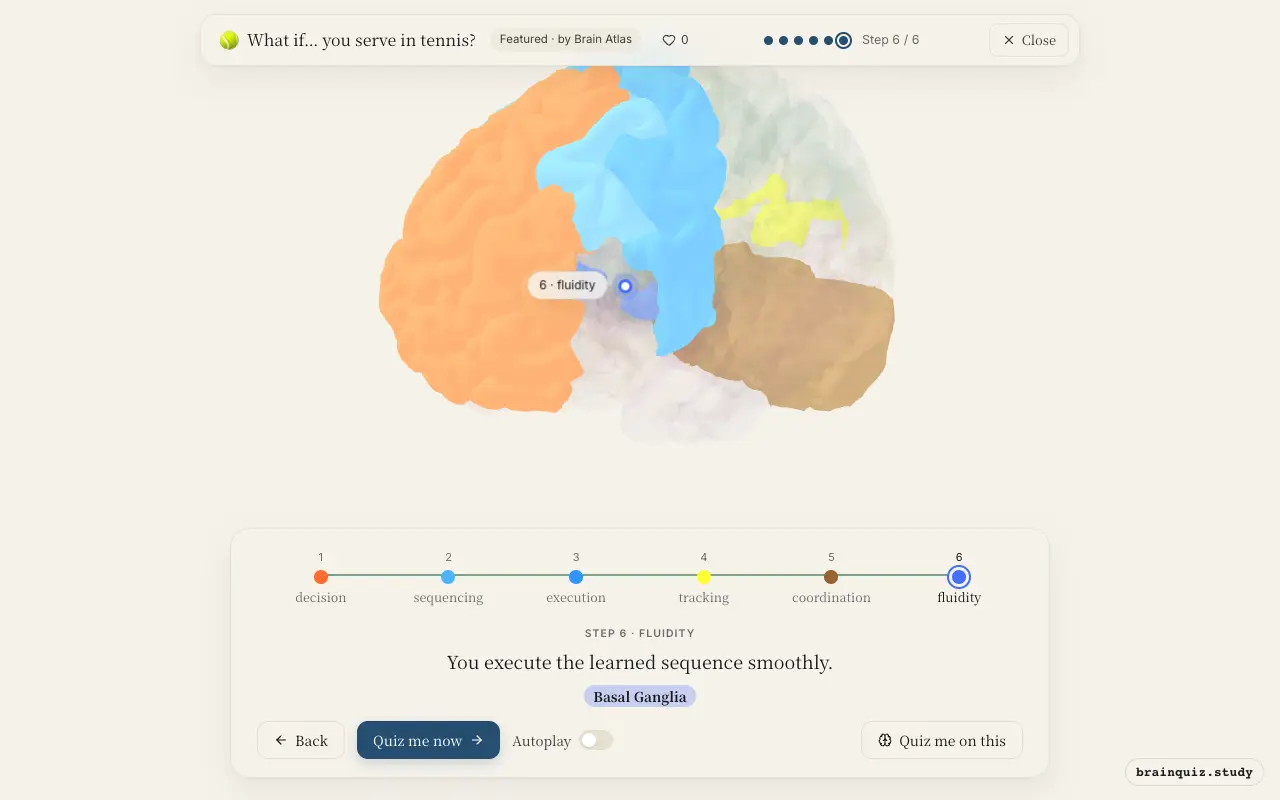
Watch it light up on the 3D brain
Brain regions that light up
| Order | Region | What it does here |
|---|---|---|
| 1 | Prefrontal Cortex | You choose where to serve and plan strategy. |
| 2 | Premotor Cortex | You prepare the toss and swing pattern. |
| 3 | Motor Cortex | You send commands to muscles for toss and swing. |
| 4 | Visual Cortex | You track the tossed ball to time the swing. |
| 5 | Cerebellum | You fine-tune movement, balance, and timing. |
| 6 | Basal Ganglia | You execute the learned sequence smoothly. |
1. Prefrontal Cortex: decision
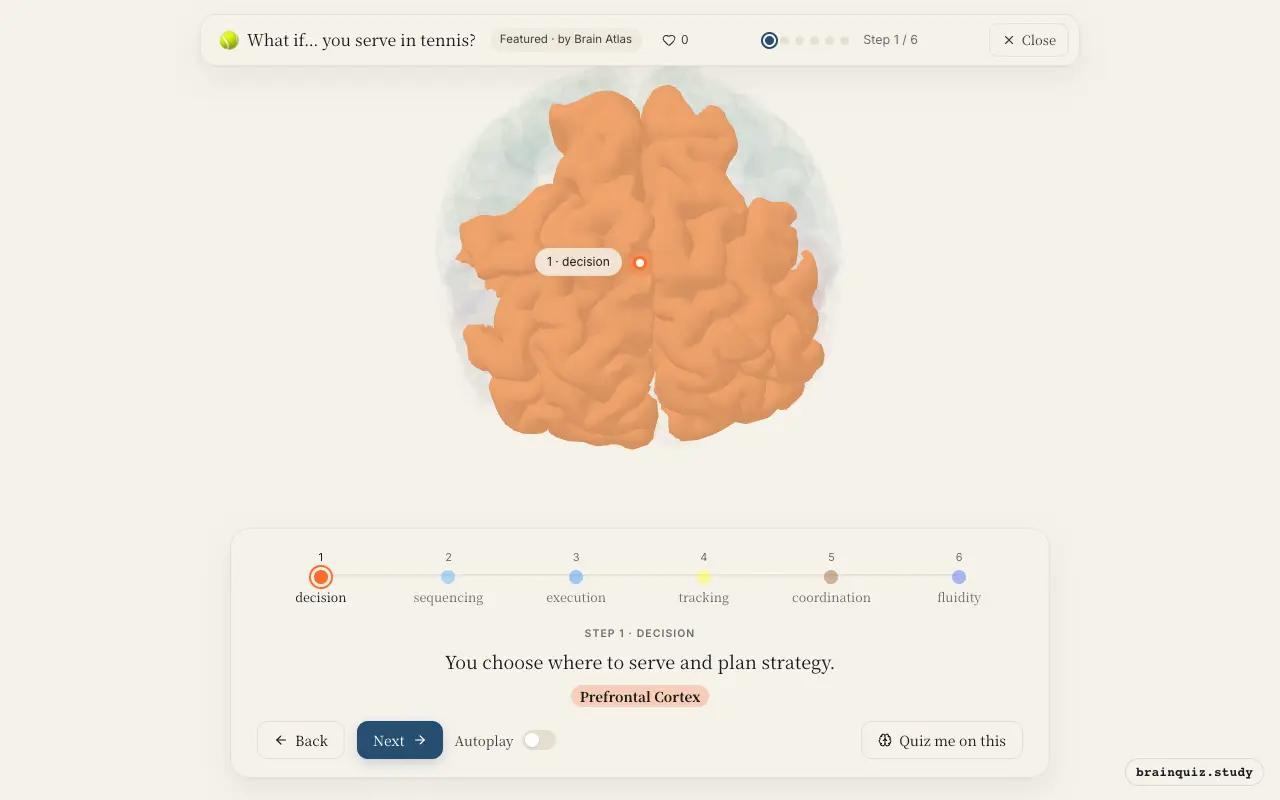
The prefrontal cortex integrates game context, opponent positioning, and past experience to decide the serve type and placement. It functions as the executive, issuing the command to begin the movement sequence. This region is critical for executive functions like planning and decision-making, setting the goal for the serve.
2. Premotor Cortex: sequencing
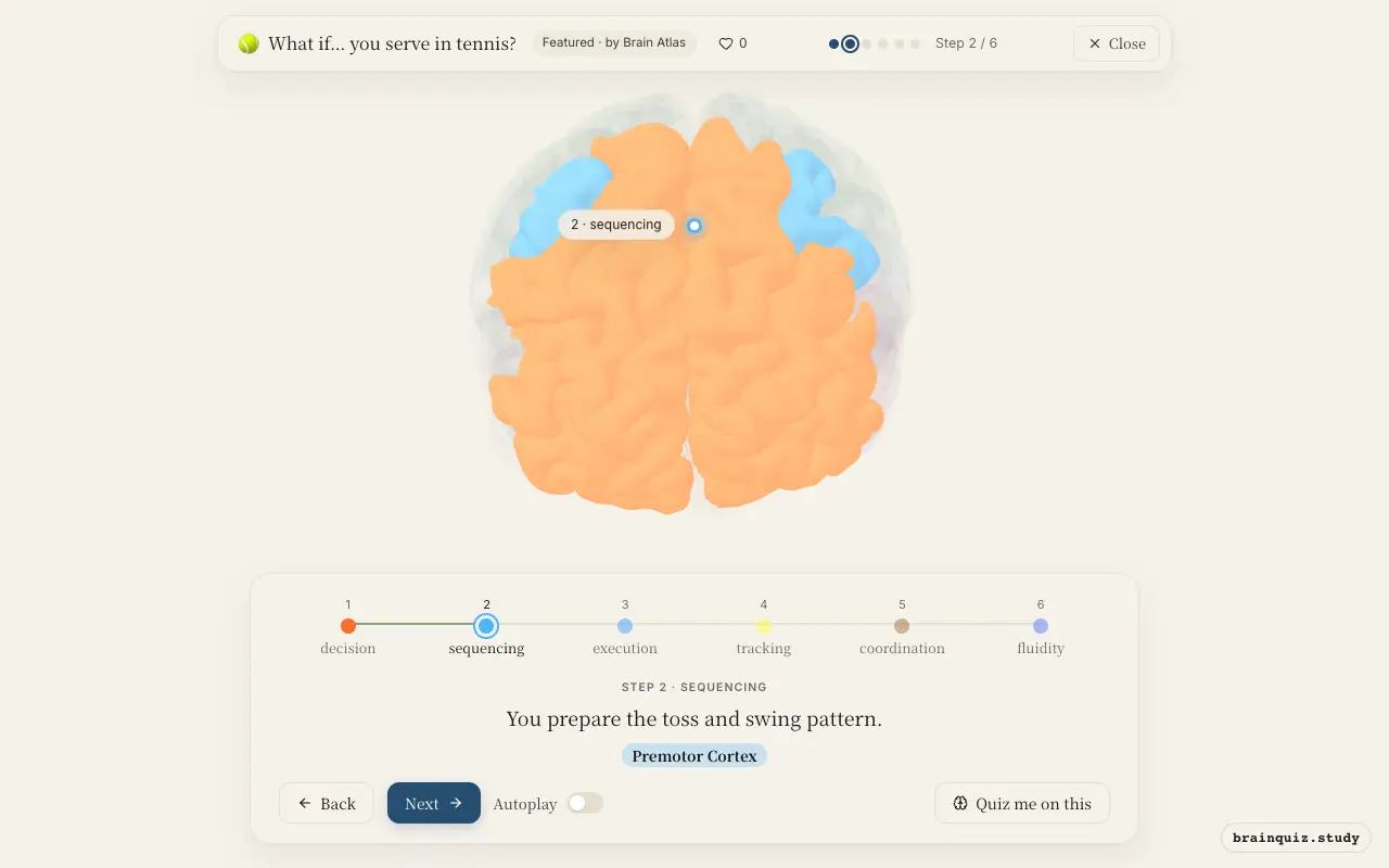
The premotor cortex organizes the temporal order of muscle activations for the serve. It receives input from prefrontal cortex and sensory areas to construct a motor program that specifies the toss, backswing, and forward swing. This region is active before movement begins, ensuring the sequence is ready for execution.
3. Motor Cortex: execution
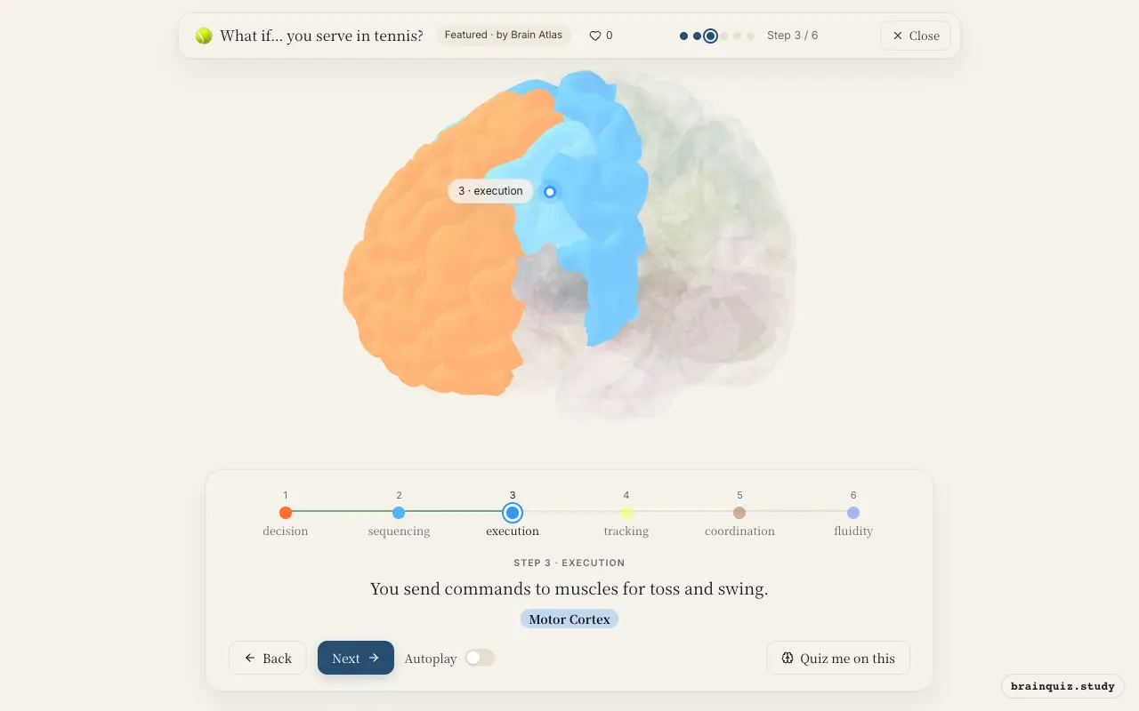
The primary motor cortex sends signals down the corticospinal tract to activate muscles in the arm, shoulder, and trunk. It initiates the toss and then the swing, with neurons firing in a precise pattern to produce the movement. This region is a major output pathway for voluntary movement.
4. Visual Cortex: tracking
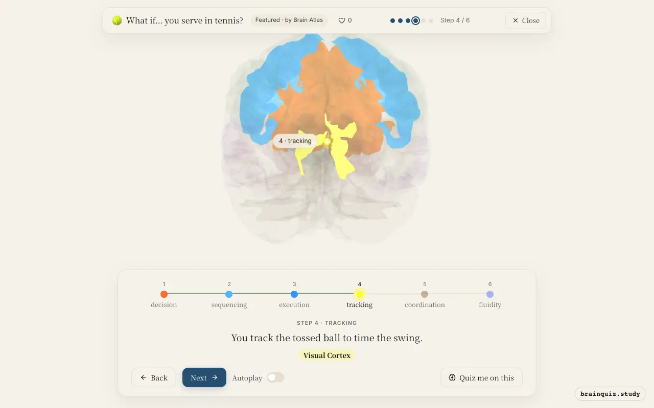
The visual cortex processes the ball's position and trajectory as it rises and falls. This information is sent to parietal and motor areas to adjust the timing of the swing, ensuring contact at the optimal point. Visual feedback is crucial for hit accuracy.
5. Cerebellum: coordination
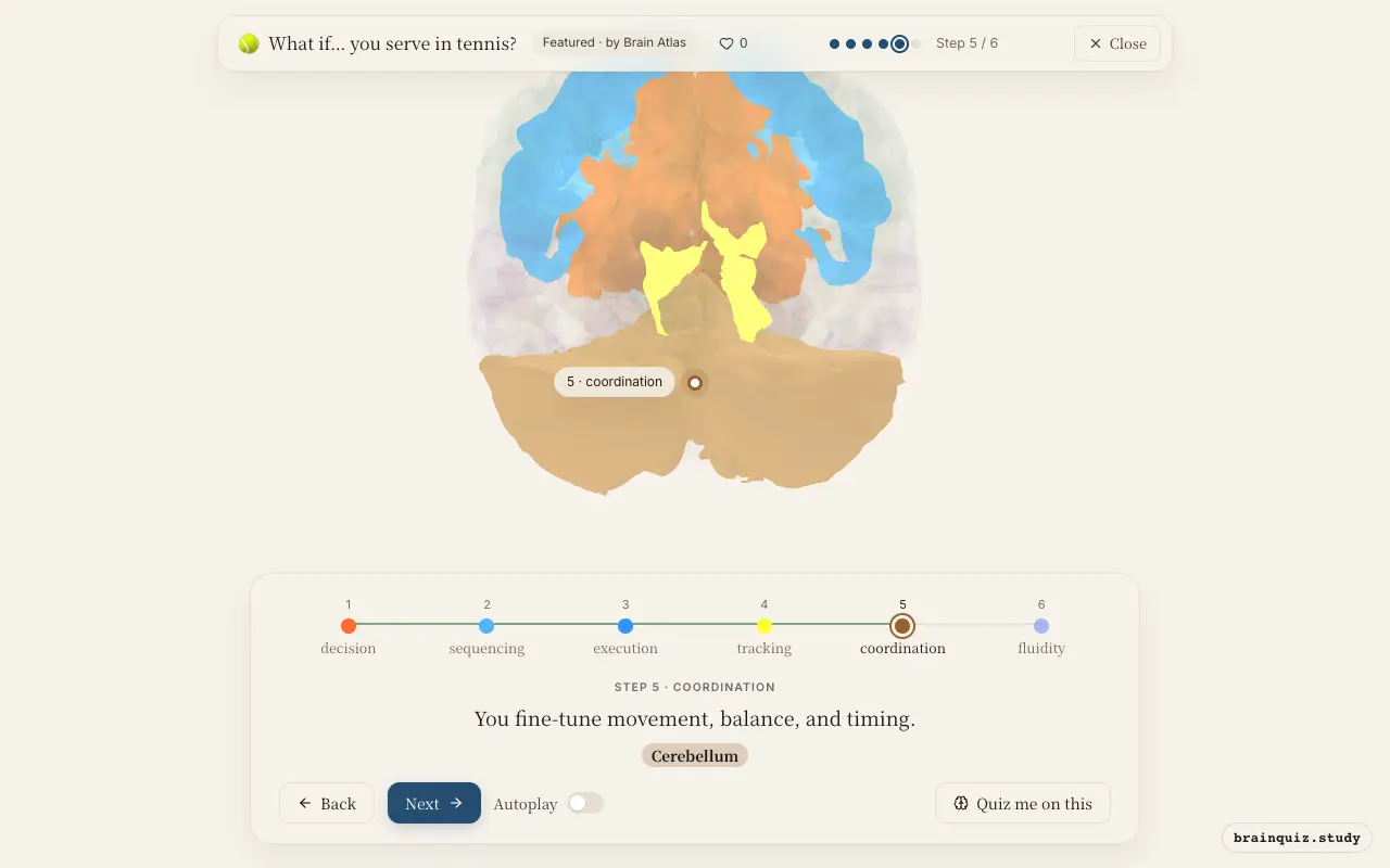
The cerebellum compares intended movement with actual sensory feedback, making real-time corrections to the swing path and body position. It ensures the serve is smooth and accurate, and it stores motor learning for future serves. It is essential for precise timing of the ball toss and contact.
6. Basal Ganglia: fluidity
The basal ganglia, particularly the striatum, facilitate the initiation and scaling of the serve by releasing the thalamus from inhibition. It suppresses unwanted movements and ensures the sequence is executed as a unified whole, reflecting well-practiced skill. This region is critical for habit formation and procedural memory.
Good to know
- The cerebellum contains more neurons than the rest of the brain combined, underscoring its importance in motor coordination.
- Basal ganglia dysfunction, such as in Parkinson's disease, leads to difficulty initiating and smoothing complex movements like a tennis serve.
- The primary motor cortex has a somatotopic organization, with a disproportionately large representation of the hand and arm for fine motor control.
Tennis serve vs. overhand throw
An overhand throw shares the basic motor sequence of a tennis serve, but the serve's self-toss demands greater cerebellar timing precision. The visual cortex tracks the tossed ball in a serve, whereas a throw often tracks a stationary or moving target. Both rely on basal ganglia for fluid execution, but the serve's learned sequence is more stereotyped.
Test yourself
Which brain region is most responsible for the initial decision of where to serve?
Which region compares intended movement with actual feedback to correct errors during the serve?
Damage to the basal ganglia would most likely impair which aspect of a tennis serve?
0 of 3 answered · 0 correct
Simplified: many regions work together at once, and this shows the main players in the order they act.