What happens in your brain when you take a placebo?
Updated
When you take a placebo, the prefrontal cortex generates positive expectations; the anterior cingulate and insula process pain signals but are downregulated; the nucleus accumbens releases dopamine in anticipation of relief; and the periaqueductal grey activates descending opioid pathways that inhibit pain.
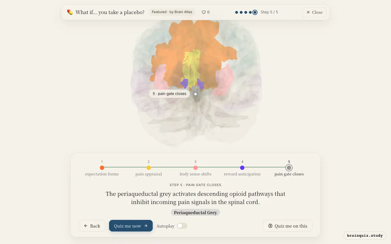
Watch it light up on the 3D brain
Brain regions that light up
| Order | Region | What it does here |
|---|---|---|
| 1 | Prefrontal Cortex | The prefrontal cortex interprets the pill as a real treatment and builds a positive expectation of relief. |
| 2 | Anterior Cingulate Cortex | The anterior cingulate cortex evaluates the placebo's meaning and reduces the emotional suffering linked to pain. |
| 3 | Insula | The insula represents internal bodily states and shows decreased pain-related activity under placebo. |
| 4 | Nucleus Accumbens | The nucleus accumbens releases dopamine as the brain anticipates relief, reinforcing the placebo response. |
| 5 | Periaqueductal Grey | The periaqueductal grey activates descending opioid pathways that inhibit incoming pain signals in the spinal cord. |
1. Prefrontal Cortex: expectation forms
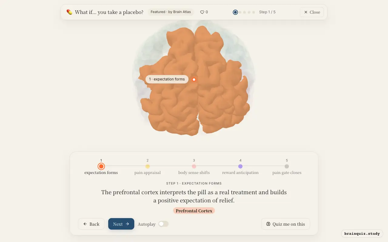
This region integrates prior experiences and social cues to generate belief that the placebo will work. Its activity correlates with the strength of expected benefit, setting the stage for downstream modulation.
2. Anterior Cingulate Cortex: pain appraisal
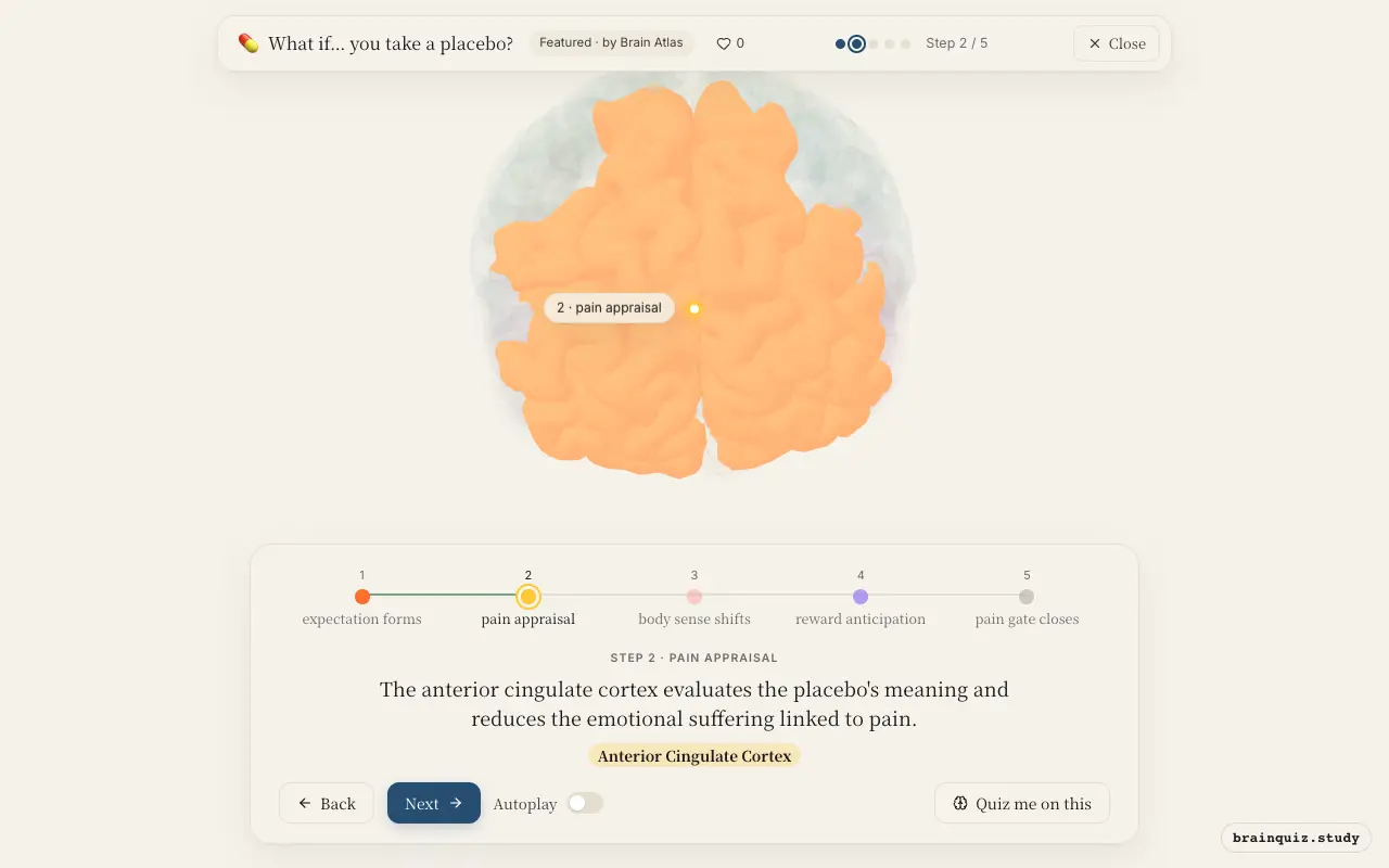
The ACC monitors conflicts between expectation and actual pain, and under placebo it shifts toward expectation-driven control. It also connects prefrontal signals to pain-modulatory networks, lessening pain unpleasantness.
3. Insula: body sense shifts
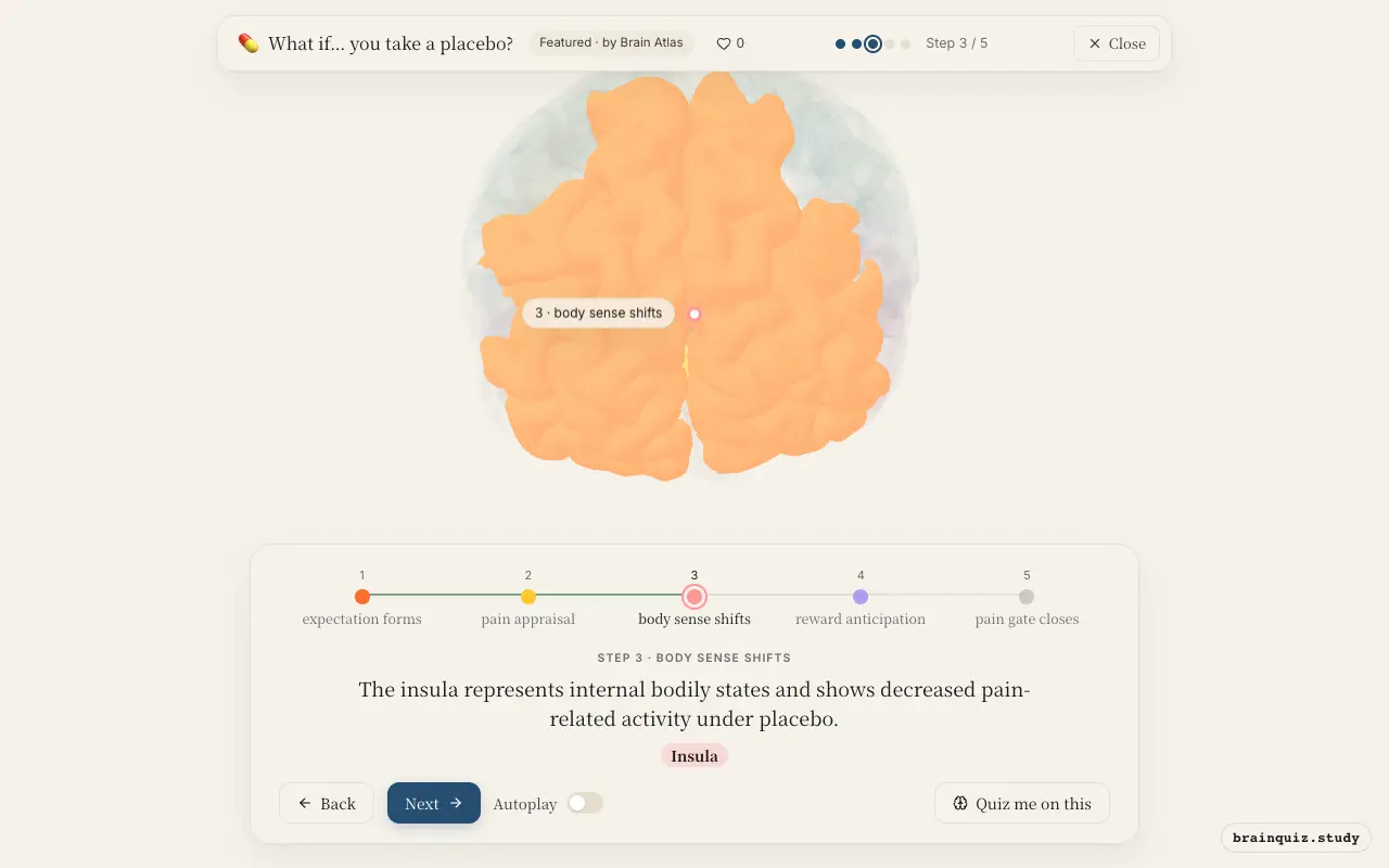
The insula normally tracks pain intensity and interoceptive awareness, but placebo-induced expectations dampen its response to painful stimuli. This reduction is thought to reflect a genuine change in perceived pain, not just reporting bias.
4. Nucleus Accumbens: reward anticipation
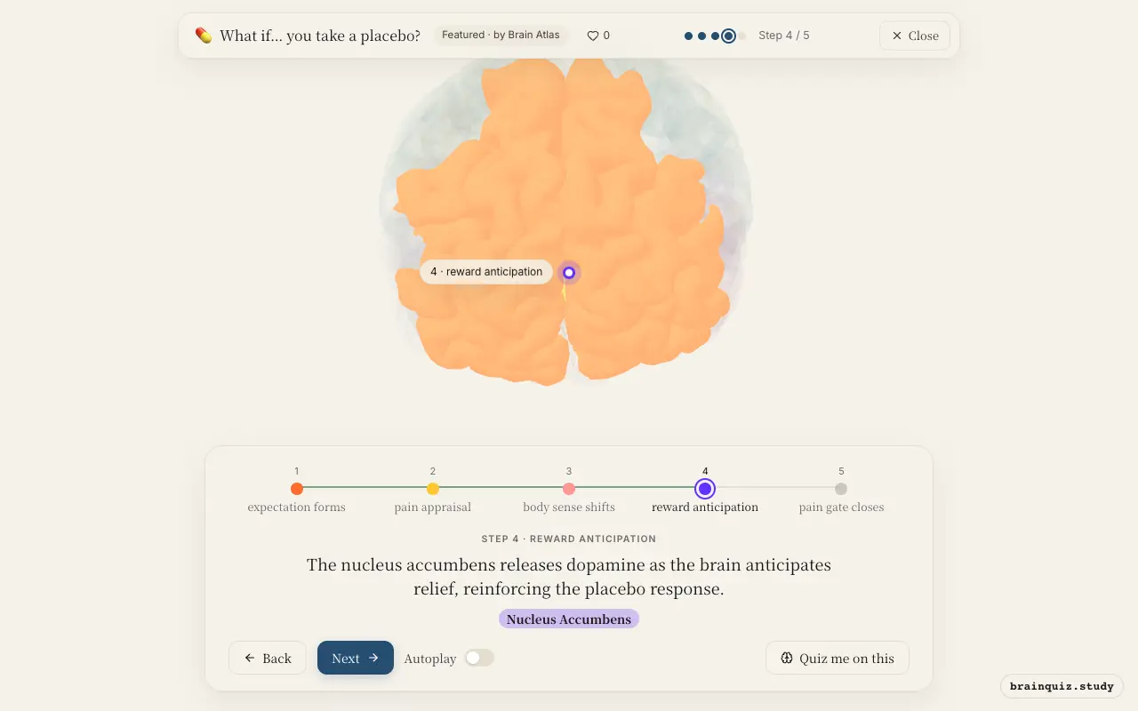
Dopamine in this reward hub signals the expectation of a positive outcome, which strengthens learning and boosts motivation. This neurochemical surge is linked to the placebo effect's ability to improve symptoms beyond pain, such as mood.
5. Periaqueductal Grey: pain gate closes
This midbrain region is a key node for endogenous analgesia, releasing opioid peptides that suppress nociceptive transmission. Placebo-induced activation here explains why opioid blockers like naloxone can abolish placebo analgesia.
Good to know
- Placebo analgesia can be blocked by naloxone, an opioid receptor antagonist, demonstrating the involvement of endogenous opioids.
- Brain imaging studies show that placebo effects are stronger when individuals have high expectations and prior successful treatment experiences.
- The placebo response is not 'all in the mind' but involves measurable changes in brain activity and neurotransmitter release.
Placebo vs nocebo
Both rely on expectation, but placebo engages prefrontal and reward circuits to inhibit pain via endogenous opioids, whereas nocebo activates anxiety and fear circuits like the amygdala, amplifying pain. The same brain networks can produce opposite outcomes based on the valence of expectation.
Test yourself
Which brain region is most responsible for generating positive expectations about a placebo?
What neurotransmitter is released in the nucleus accumbens during placebo anticipation of relief?
Which region's activation is directly linked to the release of endogenous opioids that block pain signals?
0 of 3 answered · 0 correct
Simplified: many regions work together at once, and this shows the main players in the order they act. For learning only, not medical advice.