What happens in your brain when you feel pain?
Updated
Thalamus relays pain signals to the cortex. Somatosensory cortex localizes and grades intensity. Insula produces bodily awareness. Anterior cingulate generates emotional distress. Prefrontal cortex evaluates meaning. Periaqueductal grey dampens pain via descending pathways.
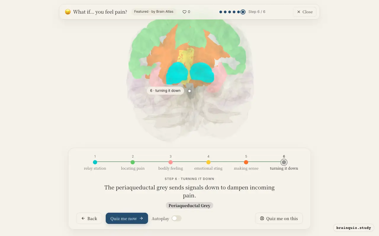
Watch it light up on the 3D brain
Brain regions that light up
| Order | Region | What it does here |
|---|---|---|
| 1 | Thalamus | Pain signals from the spinal cord arrive at the thalamus. |
| 2 | Somatosensory Cortex | The somatosensory cortex maps where the pain is and how intense. |
| 3 | Insula | The insula creates the feeling that the pain is happening to you. |
| 4 | Anterior Cingulate Cortex | The anterior cingulate adds the emotional suffering and urge to escape. |
| 5 | Prefrontal Cortex | The prefrontal cortex evaluates the pain's meaning and plans a response. |
| 6 | Periaqueductal Grey | The periaqueductal grey sends signals down to dampen incoming pain. |
1. Thalamus: relay station
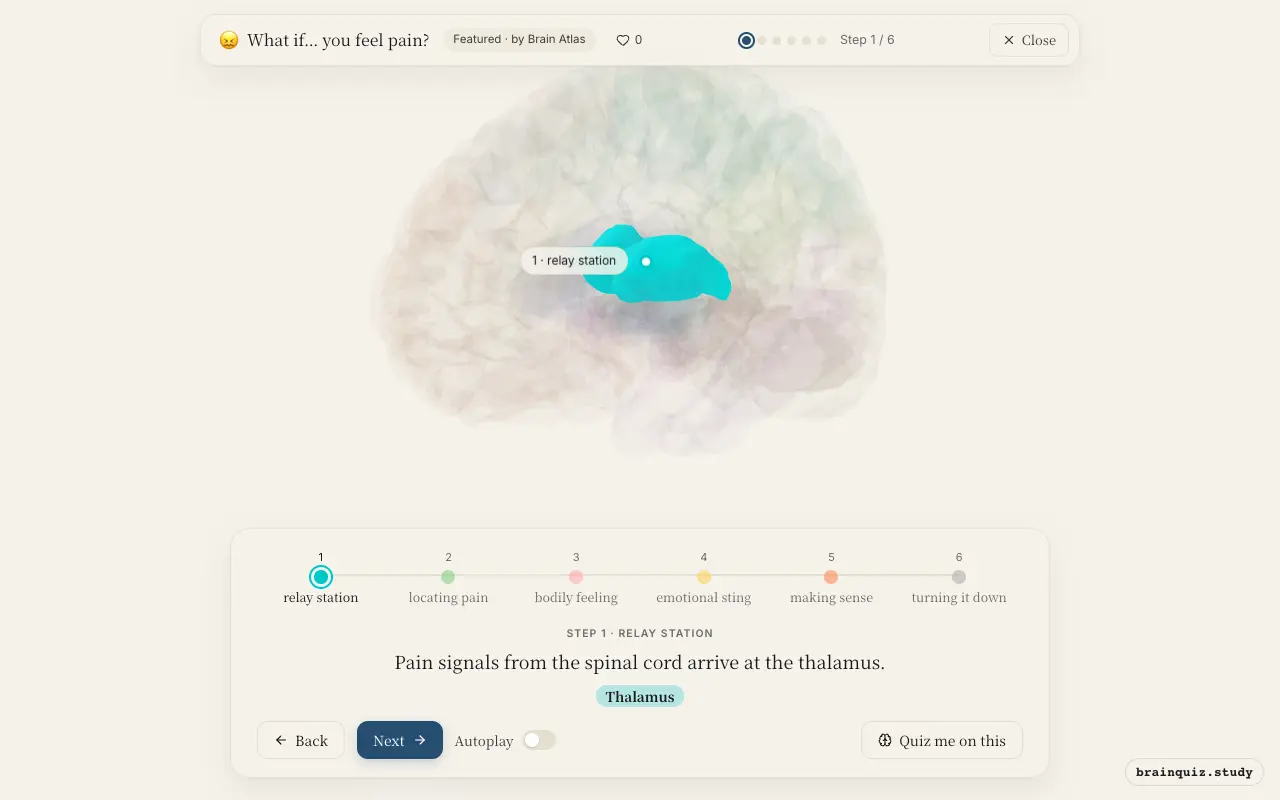
The thalamus acts as the brain's main sensory relay. Specific nuclei, such as the ventral posterior nucleus, receive nociceptive input from the spinothalamic tract and project to cortical areas. It filters and forwards signals to both sensory and limbic regions.
2. Somatosensory Cortex: locating pain
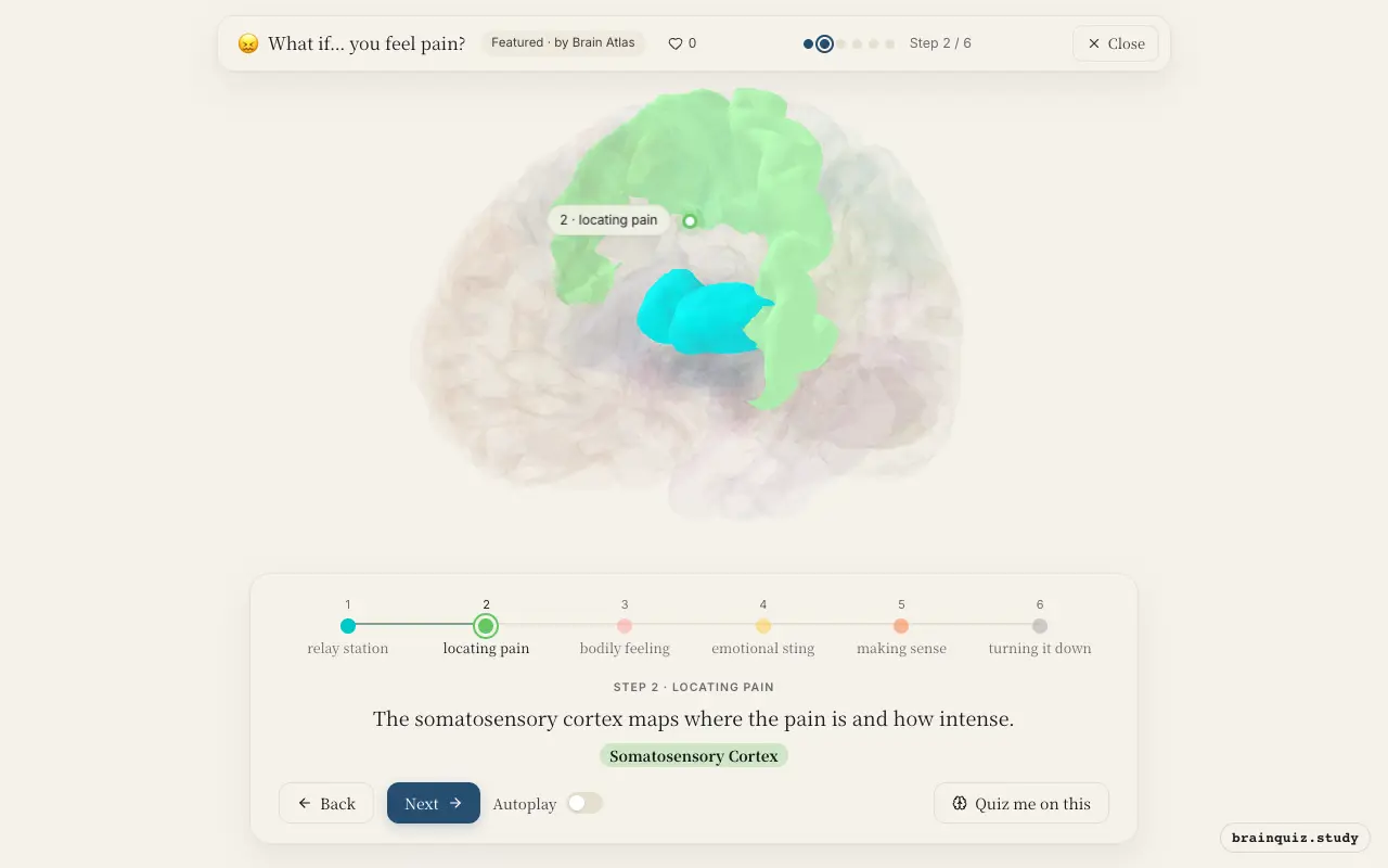
Primary somatosensory cortex (S1) receives thalamic input and encodes the sensory-discriminative aspects of pain: location, intensity, and quality. It uses a somatotopic map, so the area activated corresponds to the body part injured.
3. Insula: bodily feeling
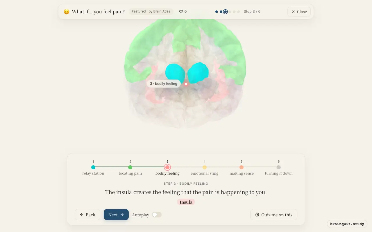
The insula integrates interoceptive signals from the body, giving rise to the subjective awareness of pain as an internal state. It is thought to be crucial for the "feeling" of pain, contributing to its unpleasantness and urgency.
4. Anterior Cingulate Cortex: emotional sting
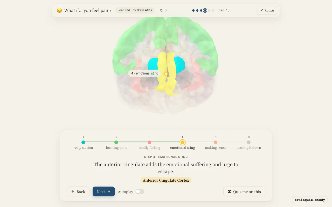
The anterior cingulate cortex (ACC) processes the affective-motivational dimension of pain. It is associated with the distress, fear, and desire to withdraw from the painful stimulus. Lesions here can reduce the suffering while leaving sensory discriminative abilities intact.
5. Prefrontal Cortex: making sense
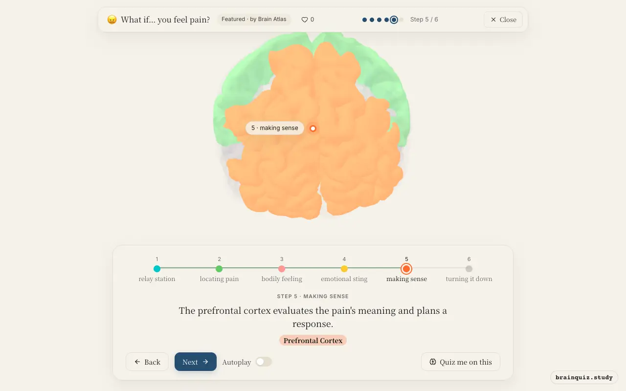
The prefrontal cortex, especially dorsolateral and ventromedial regions, appraises the pain in context: is it dangerous, controllable, or familiar? It can modulate emotional reactions and direct attention, influencing how much pain is felt and coping strategies.
6. Periaqueductal Grey: turning it down
The periaqueductal grey (PAG) is a key node in the descending pain modulation system. It receives input from the ACC and other regions and projects to brainstem nuclei that inhibit nociceptive transmission in the spinal cord, reducing pain intensity.
Good to know
- The placebo effect involves activation of the prefrontal cortex and periaqueductal grey, which can inhibit pain via endogenous opioids.
- The anterior cingulate cortex is sometimes targeted in cingulotomy, a neurosurgical procedure for intractable pain that reduces the emotional suffering.
- The insula and anterior cingulate are part of the "salience network," which detects and responds to behaviorally relevant stimuli, including pain.
Physical pain vs social pain
Physical pain activates somatosensory cortex for localization, while social pain (e.g., exclusion) does not; both activate insula and anterior cingulate, suggesting shared circuitry for distress. The periaqueductal grey may be more specific to physical pain modulation.
Test yourself
Which brain region is primarily responsible for localizing where on the body a painful stimulus occurs?
Damage to which region would most likely reduce the emotional suffering associated with pain while leaving the ability to locate it intact?
The descending pain modulation system, which can inhibit pain signals at the spinal cord, is centered in which structure?
0 of 3 answered · 0 correct
Simplified: many regions work together at once, and this shows the main players in the order they act.