What happens in your brain when you get a spinal cord injury?
Updated
A spinal cord injury severs axon tracts in the spinal cord, causing autonomic dysregulation from brainstem pathways, loss of relay in the thalamus, deafferentation of the somatosensory cortex, and disconnect of motor cortex commands, leading to paralysis and numbness below the lesion.
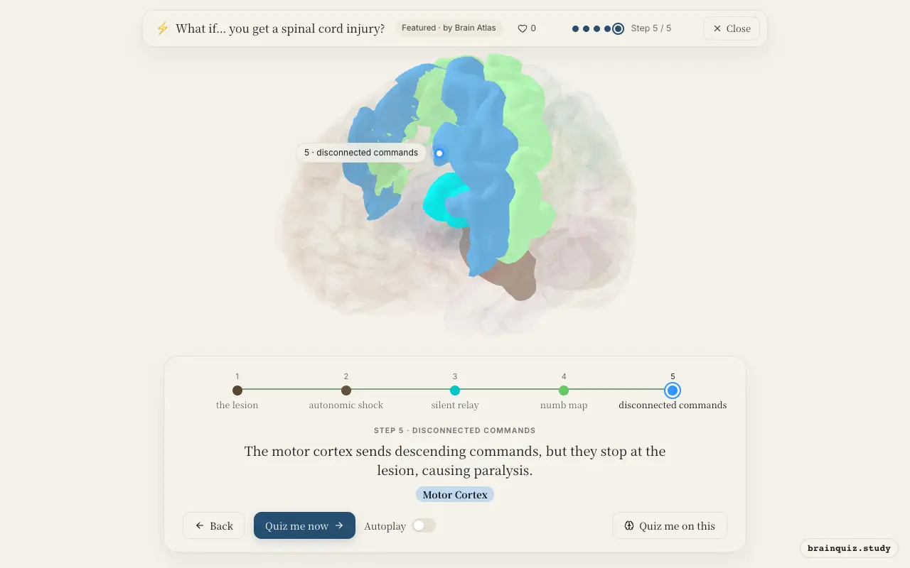
Watch it light up on the 3D brain
Brain regions that light up
| Order | Region | What it does here |
|---|---|---|
| 1 | Spinal Cord | The spinal cord is damaged, severing long axon tracts that carry sensory and motor signals. |
| 2 | Brainstem | The brainstem loses normal input and responds with altered autonomic control, risking neurogenic shock. |
| 3 | Thalamus | The thalamus stops receiving somatosensory signals from below the injury, so it cannot forward them to the cortex. |
| 4 | Somatosensory Cortex | The somatosensory cortex loses sensory input for areas below the injury, producing immediate numbness. |
| 5 | Motor Cortex | The motor cortex sends descending commands, but they stop at the lesion, causing paralysis. |
1. Spinal Cord: the lesion
The spinal cord contains ascending pathways (spinothalamic, dorsal columns) and descending pathways (corticospinal). Injury interrupts these fibers, cutting off communication between the brain and body below the lesion.
2. Brainstem: autonomic shock
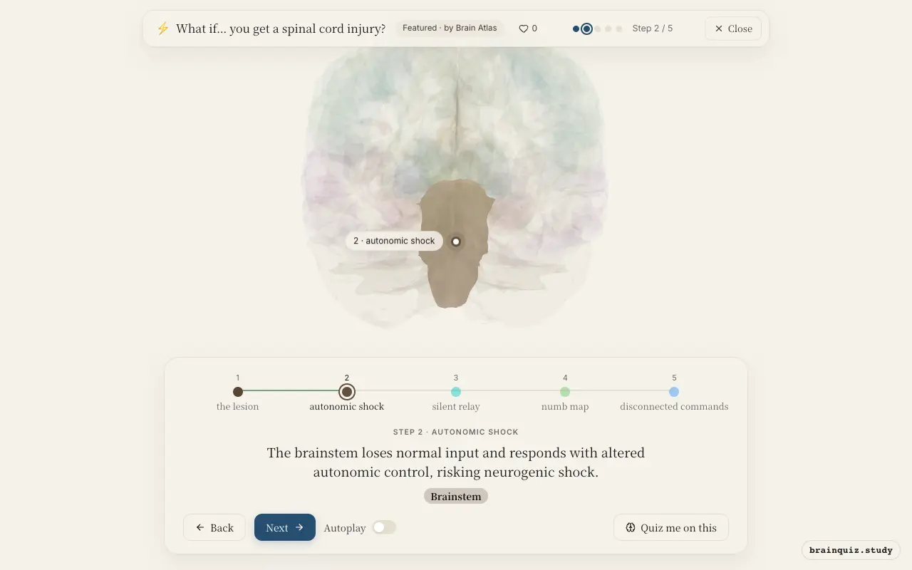
Brainstem centers regulate heart rate and blood pressure via sympathetic pathways. Loss of descending sympathetic signals to the body below the injury can cause vasodilation and low blood pressure.
3. Thalamus: silent relay
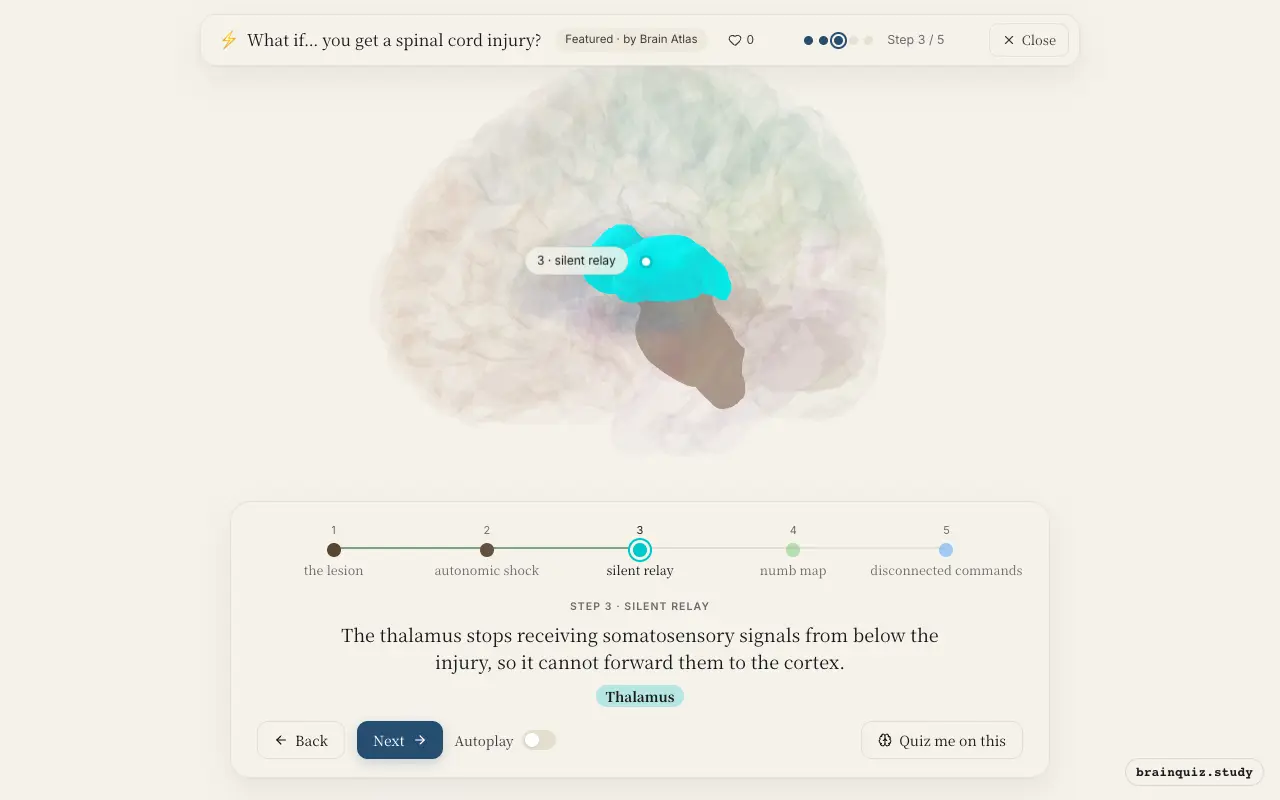
The ventral posterior nucleus of the thalamus normally relays touch and pain to the somatosensory cortex. Without input, its neurons become less active, leading to sensory loss.
4. Somatosensory Cortex: numb map
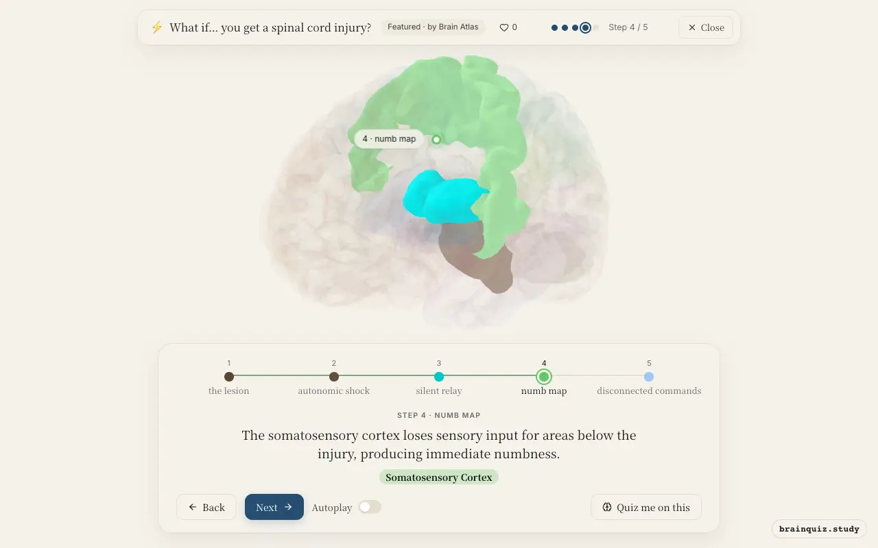
The postcentral gyrus has a somatotopic map. Deafferentation causes the corresponding cortical region to be silenced; over time, neighboring regions may invade the unused cortex.
5. Motor Cortex: disconnected commands
Primary motor cortex neurons in layer V project via the corticospinal tract. Although the cortex may still fire during attempted movement, signals cannot reach lower motor neurons below the injury.
Good to know
- The dorsal columns carry fine touch and proprioception; their interruption causes loss of discriminative sensation below the injury.
- Central neuropathic pain, common after spinal cord injury, is associated with changes in the anterior cingulate cortex and insula.
- Spinal shock causes a temporary loss of all reflexes below the lesion, but spasticity often develops later.
SCI vs cortical stroke
In a cortical stroke, the motor cortex itself is damaged, so no commands are generated. In spinal cord injury, the motor cortex often remains active and attempts to send signals, but they are blocked at the lesion. This distinction is key for brain-computer interface approaches.
Test yourself
Which brain region loses its sensory input map below a spinal cord injury?
What happens to motor cortex activity during attempted movement after spinal cord injury?
Which region is primarily responsible for the autonomic dysregulation seen in acute spinal cord injury?
0 of 3 answered · 0 correct
Simplified: many regions work together at once, and this shows the main players in the order they act. For learning only, not medical advice.