What happens in your brain when you learn to juggle?
Updated
Visual cortex tracks the balls, parietal cortex integrates spatial positions, premotor cortex plans throws, motor cortex executes commands, and cerebellum corrects errors in real time.
Watch it light up on the 3D brain
Brain regions that light up
| Order | Region | What it does here |
|---|---|---|
| 1 | Visual Cortex | The primary visual cortex processes ball positions and motion. |
| 2 | Parietal Cortex | The parietal cortex integrates visual information into a spatial map of the scene. |
| 3 | Premotor Cortex | The premotor cortex prepares the sequence of arm and hand movements. |
| 4 | Motor Cortex | The primary motor cortex sends commands to the spinal cord to move your arms and hands. |
| 5 | Cerebellum | The cerebellum compares intended movements with actual outcomes and fine-tunes motor output. |
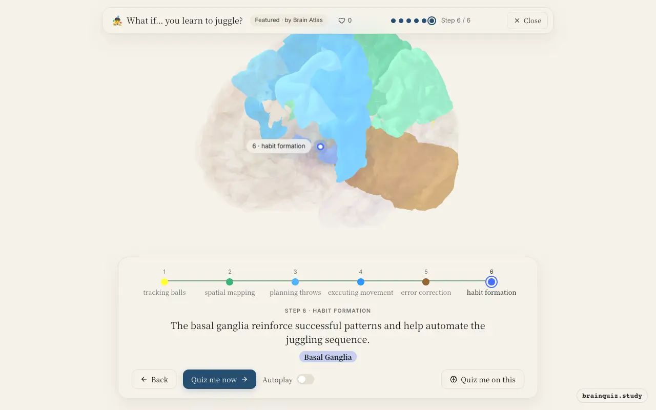
| 6 | Basal Ganglia | The basal ganglia reinforce successful patterns and help automate the juggling sequence. |
1. Visual Cortex: tracking balls
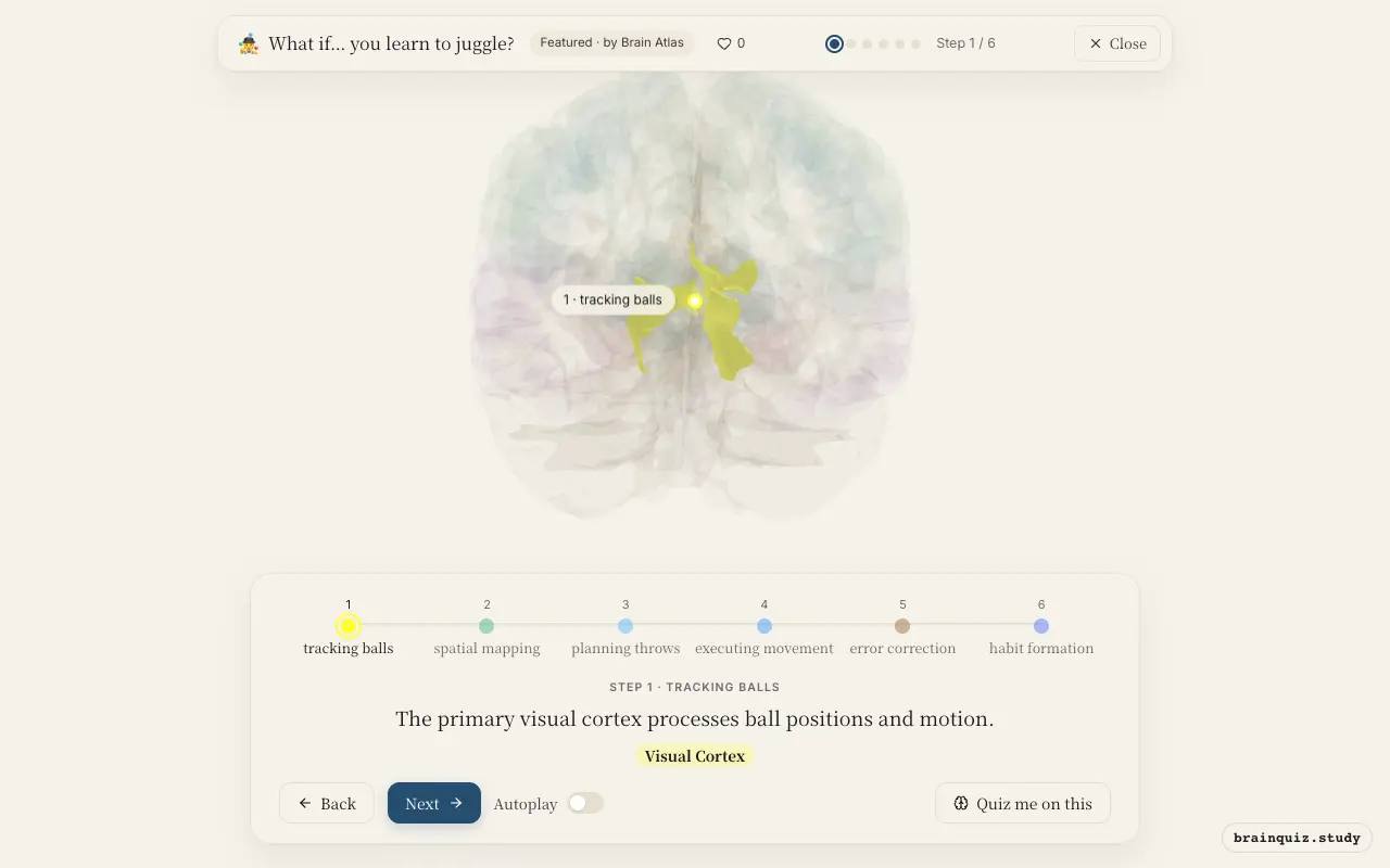
As your eyes follow the flying balls, the visual cortex in the occipital lobe decodes edges, movement, and orientation. This raw visual stream is the first step in knowing where each ball is at any instant, which is essential for planning catch and throw trajectories.
2. Parietal Cortex: spatial mapping
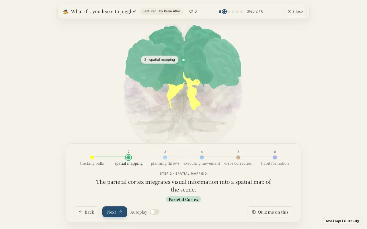
The posterior parietal cortex combines visual input with proprioceptive signals from your arms, creating a dynamic representation of ball positions relative to your body. This spatial awareness allows you to judge distances and timing, guiding your hands toward the correct catch points.
3. Premotor Cortex: planning throws
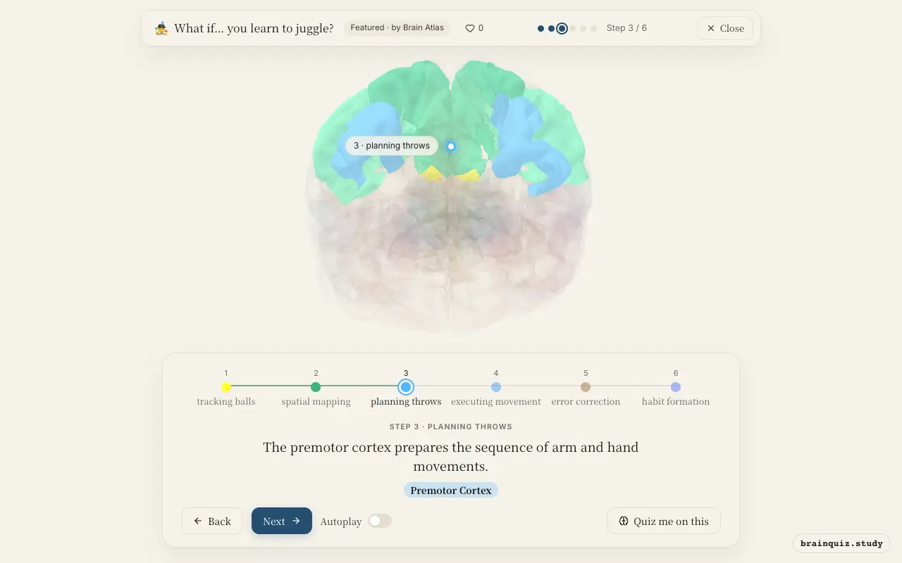
Before your muscles fire, the premotor cortex organizes the motor program for the next throw. It works with the parietal cortex to translate spatial goals into a series of muscle commands, ensuring that each toss follows the desired arc and rhythm.
4. Motor Cortex: executing movement
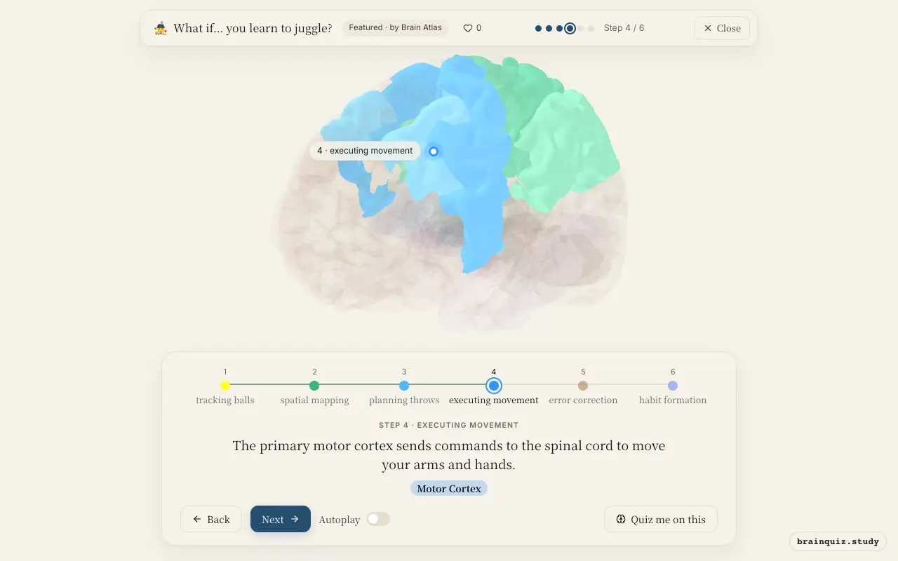
Once the premotor cortex has a plan, the motor cortex activates the upper motor neurons that project to lower motor neurons in the spinal cord. These signals cause the precise muscle contractions needed to throw and catch, adjusting force and timing on the fly.
5. Cerebellum: error correction
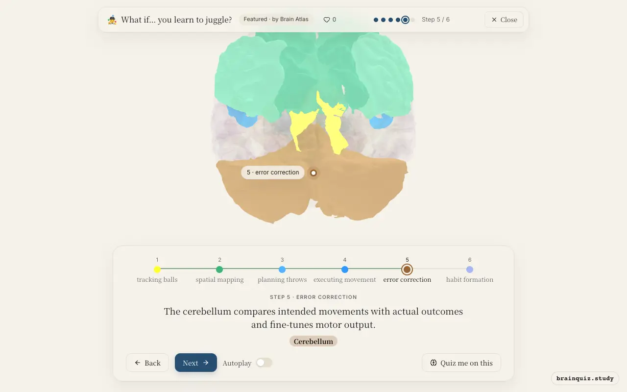
The cerebellum receives a copy of the motor plan and sensory feedback from your limbs. By detecting mismatches between expected and actual trajectories, it sends corrective signals back to the motor cortex, smoothing your movements and improving accuracy with each repetition.
6. Basal Ganglia: habit formation
As practice continues, the basal ganglia—especially the striatum—evaluate the outcomes of your throws. Dopamine signals reward successful catches, strengthening the neural pathways for efficient movement, gradually shifting juggling from effortful control to automatic habit.
Good to know
- The cerebellum contains more than half of all neurons in the brain, reflecting its role in fine motor coordination and learning.
- Juggling practice can increase grey matter density in brain areas involved in visual motion processing, as shown in some longitudinal studies.
- Motor skill learning typically shows a shift from prefrontal and parietal control to subcortical structures like the basal ganglia and cerebellum as performance becomes automatic.
Learning vs. Performing a Juggling Routine
During early learning, prefrontal and parietal regions are highly active as you consciously plan and monitor each throw. Once mastered, activity shifts to the basal ganglia and cerebellum, allowing the sequence to run automatically with minimal cortical effort, freeing attention for other tasks.
Test yourself
Which brain region is primarily responsible for error correction during motor learning?
What role does the parietal cortex play when learning to juggle?
Which structure helps automate juggling movements after extensive practice?
0 of 3 answered · 0 correct
Simplified: many regions work together at once, and this shows the main players in the order they act.