What happens in your brain when you see in the dark?
Updated
Seeing in the dark activates the thalamus to relay dim signals, the visual cortex for edge detection, the occipital cortex for motion integration, the temporal cortex for object recognition, and the parietal cortex for spatial attention.
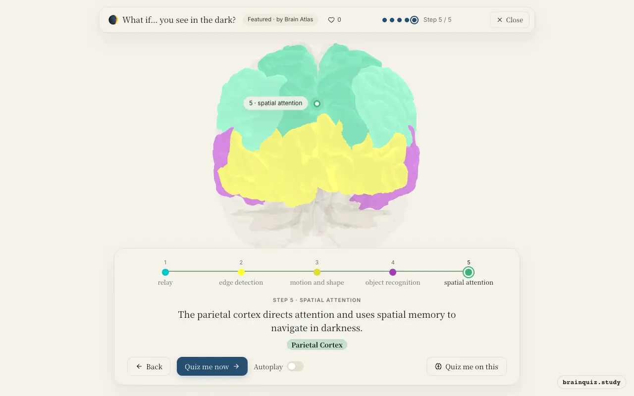
Watch it light up on the 3D brain
Brain regions that light up
| Order | Region | What it does here |
|---|---|---|
| 1 | Thalamus | The thalamus receives dim visual signals from the eyes and forwards them to the visual cortex. |
| 2 | Visual Cortex | The primary visual cortex processes basic features like edges and orientation, even in faint images. |
| 3 | Occipital Cortex | Higher visual areas integrate motion and shape information to build a coherent percept. |
| 4 | Temporal Cortex | The temporal cortex matches the processed visual input to stored object representations. |
| 5 | Parietal Cortex | The parietal cortex directs attention and uses spatial memory to navigate in darkness. |
1. Thalamus: relay
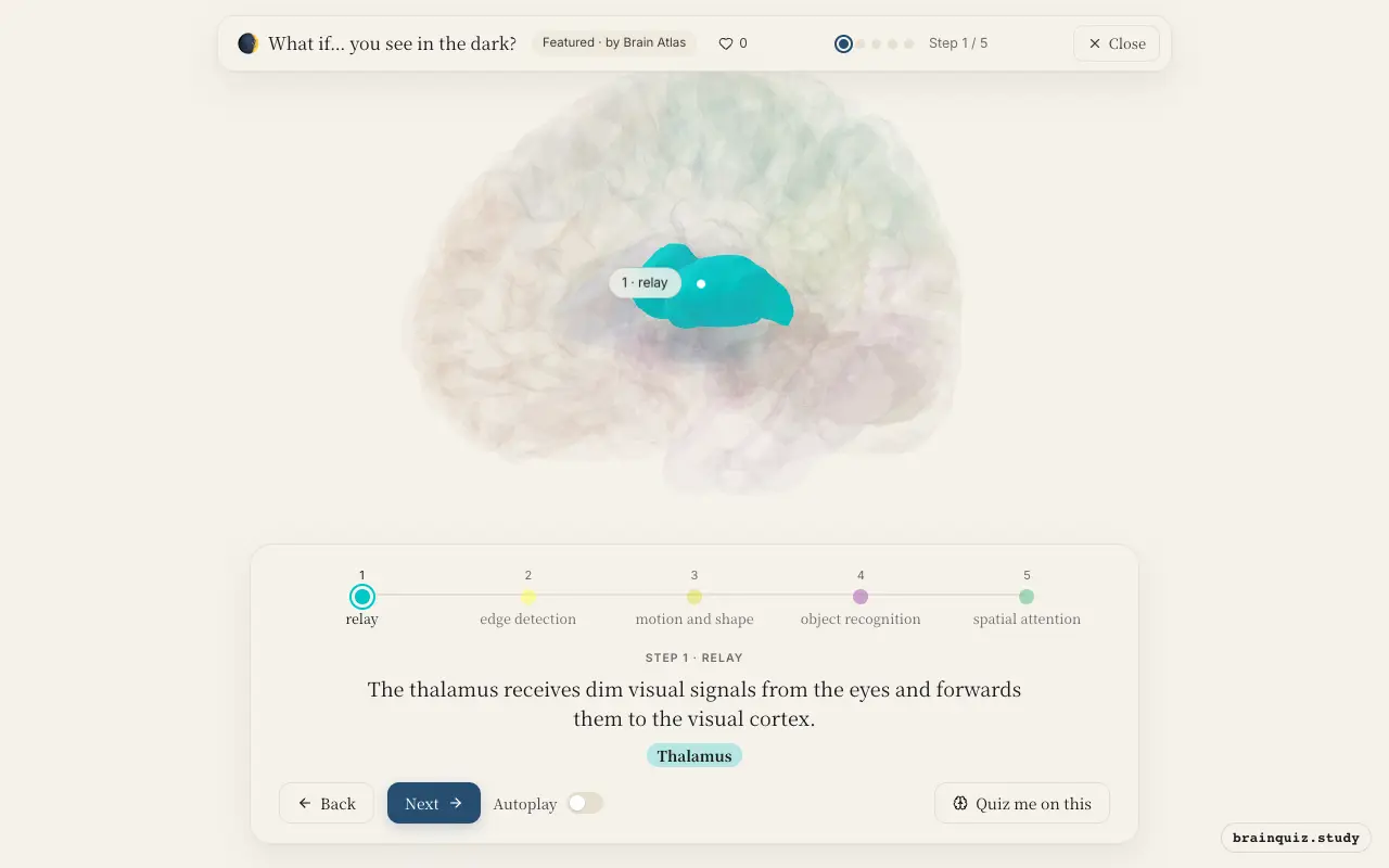
The lateral geniculate nucleus, part of the thalamus, serves as the main relay for visual information. In low light, signals from rod photoreceptors are transmitted here and then sent to the primary visual cortex, maintaining the spatial layout of the visual field.
2. Visual Cortex: edge detection
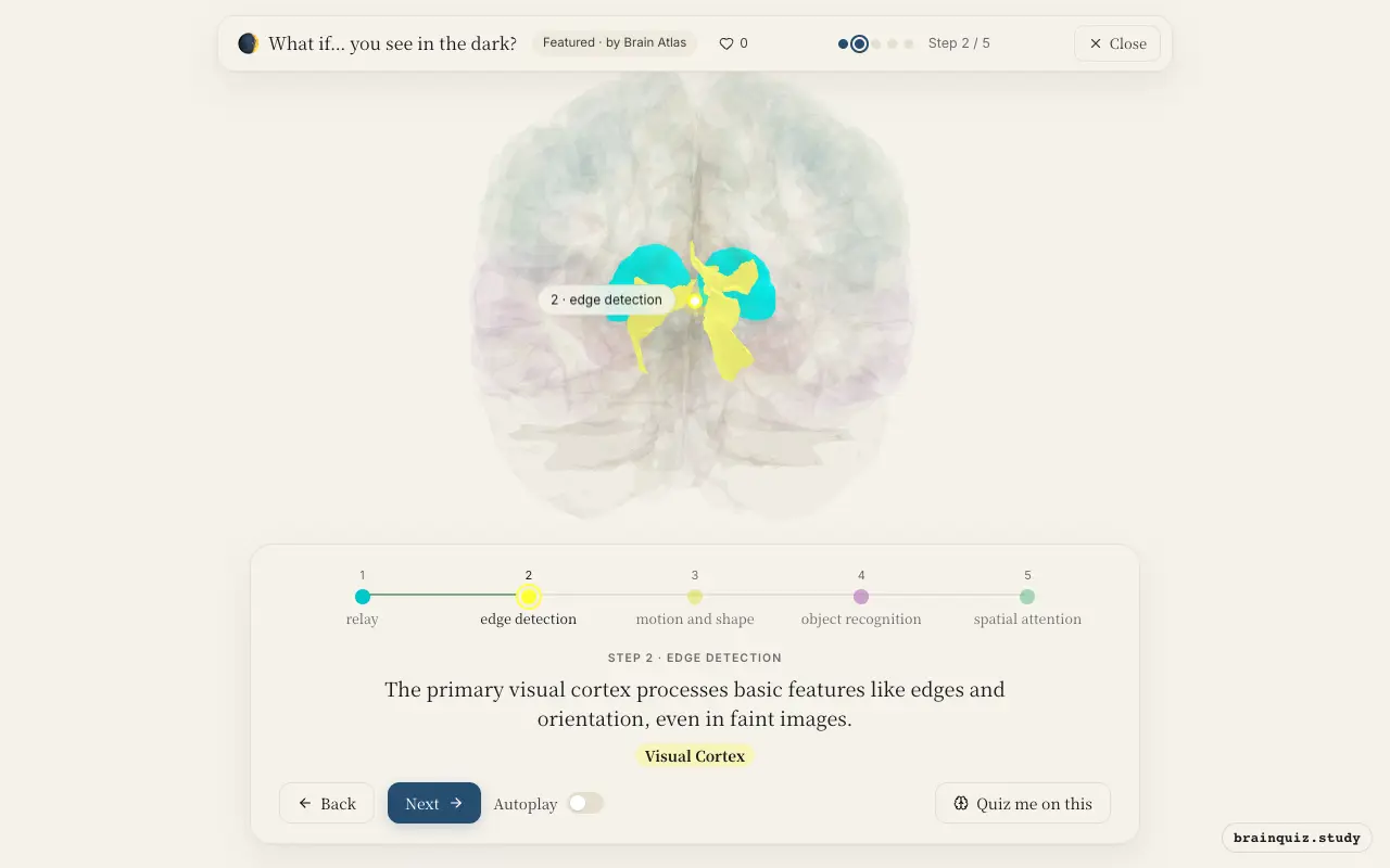
Neurons in the primary visual cortex (V1) respond to contrast and orientation, but with fewer photons, the signal-to-noise ratio is poor, so the cortex integrates over time and space to detect faint edges and contours that define shapes.
3. Occipital Cortex: motion and shape
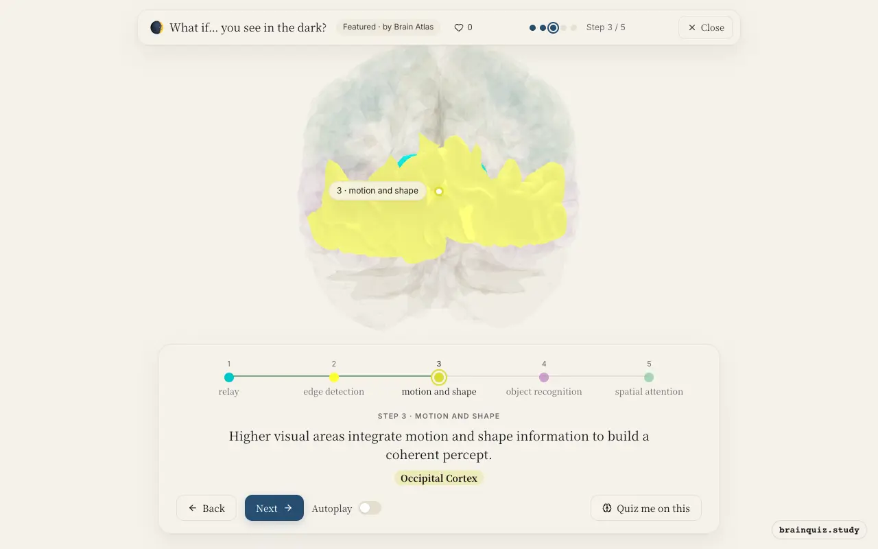
The occipital association cortex, including areas V3 and V5, combines edge and orientation information into larger contours and detects motion. Since color information is largely absent in the dark, motion and luminance boundaries become critical for perceiving moving objects.
4. Temporal Cortex: object recognition
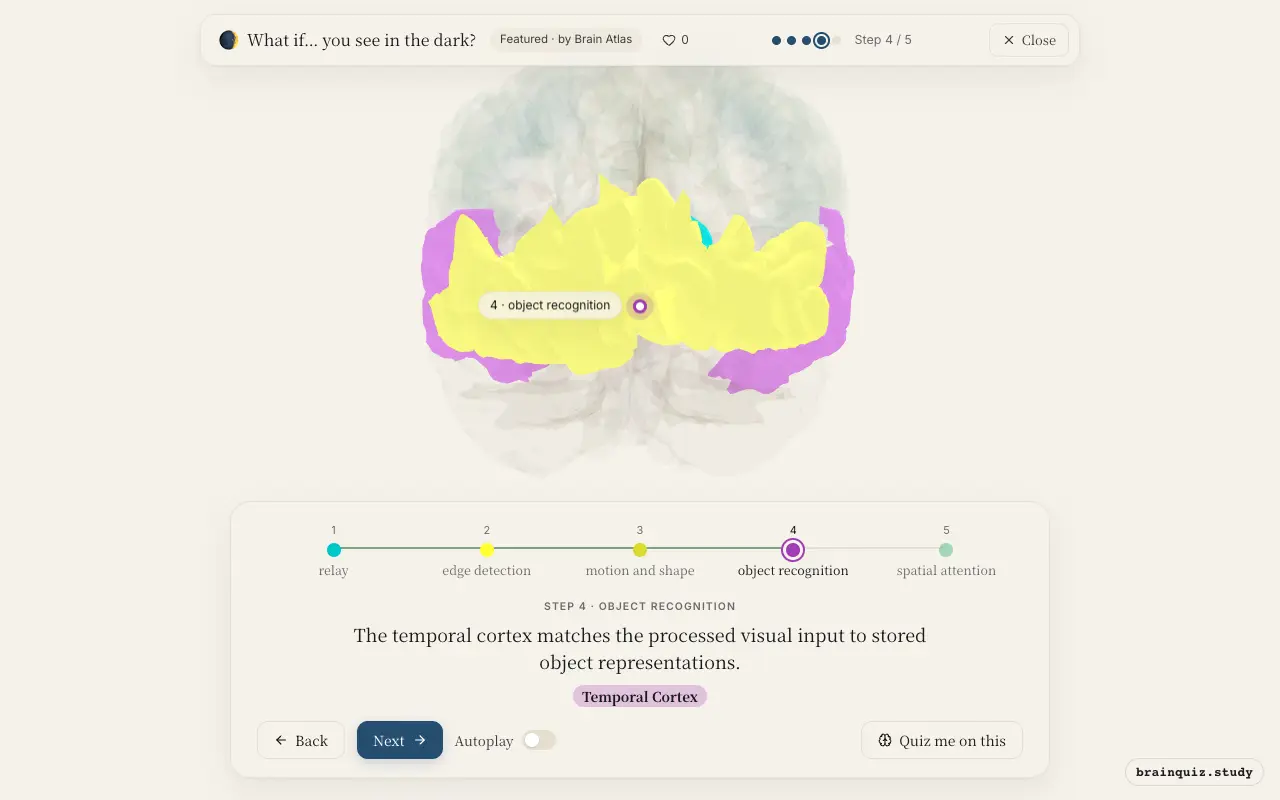
Along the ventral visual stream, the temporal cortex compares the degraded image to memory templates to identify what you are seeing. This process relies on prior knowledge and contextual cues, because the ambiguous input may not uniquely specify an object.
5. Parietal Cortex: spatial attention
The dorsal stream, including the parietal cortex, constructs a spatial map of the environment and guides eye movements and reaching actions. In darkness, this region becomes more active as you rely on memory and attentional selection to locate objects and avoid obstacles.
Good to know
- Rods in the retina are far more sensitive than cones and dominate in low light, but they do not provide color information.
- Dark adaptation, the process by which the eyes become more sensitive to dim light, can take up to 30 minutes and involves regeneration of photopigments in rods.
- Reaction times are slower in the dark because the visual system must integrate weak signals over longer periods, reducing temporal resolution.
Dark vs bright vision
In bright light, cones provide high acuity and color, so the visual cortex processes fine details and chromatic information. In the dark, rods dominate, yielding coarse, monochrome perception, and the brain relies more on motion cues and prior knowledge from the temporal cortex to interpret ambiguous scenes.
Test yourself
Which brain region first receives visual information from the eyes?
What is a key challenge for visual processing in the dark?
Which region is most involved in recognizing objects from degraded visual input?
0 of 3 answered · 0 correct
Simplified: many regions work together at once, and this shows the main players in the order they act.