What happens in your brain when you take a painkiller?
Updated
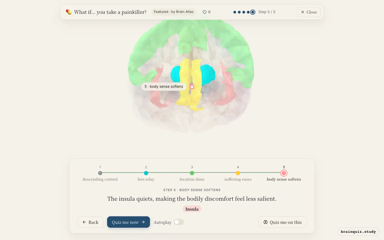
Painkillers tap into the brain's own pain-control system. The periaqueductal grey gets activated, quieting incoming pain signals; the thalamus relays less pain information; the somatosensory cortex, anterior cingulate cortex, and insula all show reduced activity, dampening the sensory, emotional, and bodily awareness of pain.
Watch it light up on the 3D brain
Brain regions that light up
| Order | Region | What it does here |
|---|---|---|
| 1 | Periaqueductal Grey | The drug activates the periaqueductal grey, the brain's built-in pain brake. |
| 2 | Thalamus | With less input from the spinal cord, the thalamus relays a weaker pain signal. |
| 3 | Somatosensory Cortex | The somatosensory cortex gets less activation, so the location and intensity of pain fade. |
| 4 | Anterior Cingulate Cortex | The anterior cingulate cortex calms, reducing the emotional suffering of pain. |
| 5 | Insula | The insula quiets, making the bodily discomfort feel less salient. |
1. Periaqueductal Grey: descending control
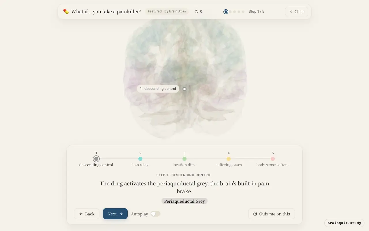
Many painkillers, especially opioids and acetaminophen, act on the periaqueductal grey (PAG) to enhance descending inhibitory pathways. The PAG sends signals down the spinal cord to suppress incoming pain messages. This is thought to be a key mechanism for central pain relief.
2. Thalamus: less relay
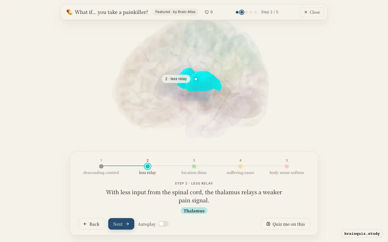
The thalamus is the brain's main sensory relay station; it receives nociceptive information from the spinothalamic tract and forwards it to cortical areas. When descending inhibition reduces spinal cord transmission, fewer pain signals reach the thalamus, so less pain information is sent to the cortex.
3. Somatosensory Cortex: location dims
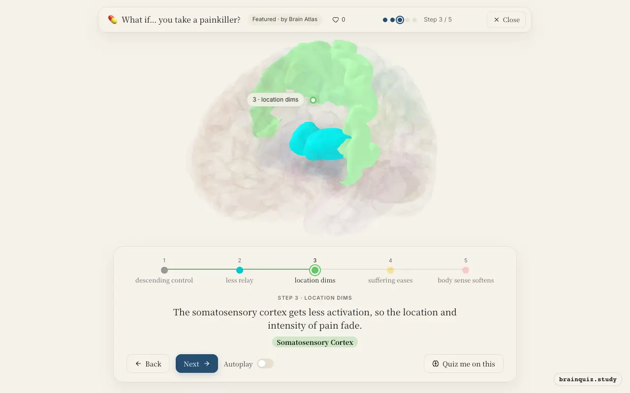
The primary somatosensory cortex processes the sensory-discriminative dimension of pain: where it is and how intense it feels. Reduced input from the thalamus means this region shows less activity, which correlates with a decrease in perceived pain intensity and localization.
4. Anterior Cingulate Cortex: suffering eases
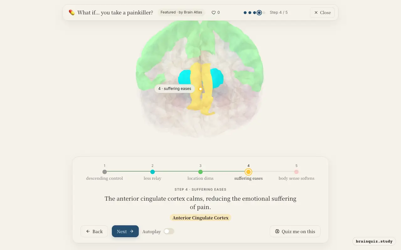
The anterior cingulate cortex (ACC) is involved in the affective-motivational component of pain, including the unpleasantness and urge to escape. Painkillers that reach the brain can dampen ACC activity directly or via reduced input, which lessens the emotional distress associated with the pain.
5. Insula: body sense softens
The insula integrates interoceptive signals and contributes to the awareness of bodily states, including pain. Decreased insular activity is seen with effective analgesia, correlating with reduced subjective awareness of the painful sensation and a sense of bodily ease.
Good to know
- Different classes of painkillers work on different molecular targets: opioids activate mu-opioid receptors, NSAIDs inhibit cyclooxygenase enzymes, and acetaminophen acts through several central mechanisms.
- The descending pain-modulation system also involves the rostral ventromedial medulla, which receives input from the periaqueductal grey and sends inhibitory signals to the spinal cord.
- Pain is a complex experience involving sensory, emotional, and cognitive components, so effective pain relief often requires reducing activity across a network of brain regions.
Painkiller vs placebo
A placebo can also reduce pain by activating similar brain regions, but it relies on expectation and endogenous opioid release rather than an external drug. With a painkiller, the effect is more consistent and often larger, though both engage the descending pain-control system.
Test yourself
Which brain region is the brain's own pain-control center that many painkillers activate?
What happens to the thalamus when a painkiller takes effect?
Which brain region is most associated with the emotional suffering component of pain?
0 of 3 answered · 0 correct
Simplified: many regions work together at once, and this shows the main players in the order they act. For learning only, not medical advice.